
Human Immune Leukoderma: Vitiligo
Overview:
There are several diseases marked by a lack of pigment in the skin that are grossly referred to as leukoderma; some are caused by an inability of melancocytes to produce melanin, while others are caused by melanocytes either not being present or being destroyed. The latter are the pathology of the phenotypically similar traits piebaldism and the disease vitiligo. Piebaldism, which is present from birth, is a lack of melanocytes in the skin, while vitiligo is a progressive disease in which the melanocytes are gradually destroyed causing unpigmented areas on the skin. The exact etiology of vitiligo is unknown, but four main theories exist to explain it: the autoimmune hypothesis, the neural hypothesis, the self-destruct hypothesis, and the growth factor defect hypothesis. It is believed that vitiligo is a polygenic trait and that a convergence theory, combining elements of different theories across a spectrum of expression is the most accurate etiology (Njoo & Westerhof 2001). Vitiligo is not a physically damaging disease; other than an increased sensitivity to UV radiation most of the disease’s effects are social and psychological, especially for dark-skinned races. There are both surgical and non-surgical treatments for vitiligo (Taneja 2002).
Picture 1. An African child exhibiting the piebald universalis phenotype-albinism. Notice children with wildtype phenotype in background. Picture from The appearance of ancient Israelites website.
 .
.
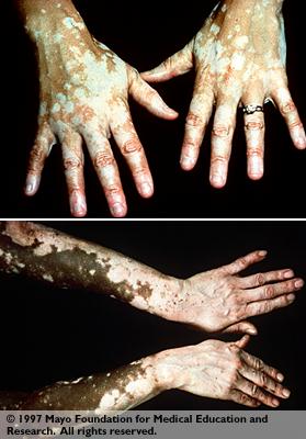
Picture 2. Two people exhibiting the vitiligo phenotype; notice wildtype pigment-producing skin is present in both pictures. Top panel is a white patient with vitiligo; the lower panel is a black patient with vitiligo. Picture from The appearance of ancient Israelites website.
Overview of Melanogenesis and Melanocytes:
The color in human hair, skin and irises is produced by the pigment melanin, which is produced by the dermal melanoycte cells. The melanocyte cells transform the peptide tyrosinase into two different forms of melanin, which then is spread throughout the dermal cells and the keratinocytes via melanosomes to darken tissue. Figure 1 shows the chemical metabolism that occurs intra-cellulary to produce melanin from the precursors phenylalanine and tyrosine; this figure is somewhat inaccurate as the end product of melanogenesis should be two different types of melanin, eumelanin and pheomelanin. Eumelanin is metabollized from DHICA and produces a brown color in hair in its intact form; pheomelanin is metabolized from 5,6-indolequione, which produces a red color in hair in its intact form. From these two slightly different forms of pigment in various degrees of structural integrity come all the differing shades of Caucasian hair (Prota 2000). In addition to coloration, melanin pigmentation in the skin also provides photoprotection from UV radiation to the skin. However, the melanocytes themselves are not immune from radiation damage: melanomas are tumors of the melanocytes, which often present themselves as discolorations owing to the pigment-producing nature of the cells (Janeway 2001).
Figure 1. Intracellular transformation of tyrosinase into pre-melanin metabolites, and finally into melanin; several of the metabolites between tyrosinase and melanin are toxic to melanocytes according to the self-destruct theory. Figure from personal website of Dr. Rodolfo Nicolaus; permission pending.
Theories for the etiology of Vitiligo
Autoimmune Theory:
There is great anecdotal evidence that an autoimmune disorder causes the destruction of melanocytes, and this theory is now generally accepted as the common cause of vitiligo. It is known vitiligo appears in conjunction with several other autoimmune disorders, such as juvenile diabetes mellitus, Addison's disease, and pernicious anemia, and additionally organ-specific antibodies can often be seen in patients with vitiligo. If the immune system raises antibodies or cytotoxic T cells to damage melanocytes, the mode of action the cells take against the melanocytes could be apoptosis induction directly against melanocytes or Ig induced complement-both are demonstrated in figure 3. Proving this theory, there is histological evidence in vitiligo patients that apoptosis is occurring in the unpigmented skin lesions: there is damage to the melanocytes and keratinocytes in these areas, and the melanocytes exhibit nuclear shrinking, vacuolization, loss of dendrites, and detachment. If antibodies do cause vitiligo, some research suggests the Ig’s may bind to tyrosinase related proteins 1 and 2, which are important for melanogenesis, instead of Ig's targeting the melanocytes directly (Huang et al. 2002).
Figure 3. Autoimmue theory of vitiligo showing both cell-mediated and humoral autoimmune responses. Figure from Expert Reviews on Molecular Medicine online; permission pending.
Neural Theory:
There is also evidence that peripheral nerve endings may secrete
a substance that is cytotoxic to melanocytes and causes their destruction. This
is supported by the segmental variety of vitiligo, which occurs in specific
dermatomes, indicating the skin is possibly only being affected by the nerves
of that specific dermatome. Additionally, vitiligo appears with certain neurological
disorders such as encephalitis, and trauma that causes peripheral nerve damage.
Nerve endings in depigmented areas were seen to produce abnormal neuropeptides
and nerve growth factors, and displayed axonal degeneration-these abnormal chemicals
may be toxic to melanocytes. Additionally, depigmented areas showed some abnormal
autonomic function, such as increased adrenergic tone, increased norepinephrine,
and an increased concentration of catecholamines. These data then suggest that
neurotransmitter release could, directly or indirectly, have an affect on melanocyte
destruction and depigmentation.
Self-Destruct Theory:
It is known that some of the intracellular pre-melanogenesis
metabolites are toxic to melanocytes, such as dopa and dopachrome. Normally
melanocytes possess cellular measures to counteract these toxic substances,
but it is believed that cells may lose the ability to counteract these toxic
metabolites and are destroyed by leakage of metabolites into the cytoplasm and
eventually cell lysis. There is evidence that points to this in that certain
hydroquinone derivatives that are similar to these intra-cellular metabolites
cause leukoderma experimentally.
Growth Factor Defect Hypothesis:
A study in the 1980's found that melanocytes in lesions from
vitiligo patients contained melanocytes, but that these cells exhibited "defective
growth and passage capacities." The researchers then noted that the growth
defects of the melanocytes were partially corrected by adding a growth factor
to their culture, additionally suggesting that growth defects may be part of
the pathology of vitiligo. In depigmented areas, cellular analysis showed that
there were melanocytes but that they grew poorly. These data suggest that, whether
a primary or secondary cause, growth defects appear to play a role in leukoderma
and vitiligo (Njoo & Westerhof 2001).
Genetic Influences:
There does appear to be a strong genetic influence in vitiligo: a positive family history has been reported in about 20% of patients and it has been found in monozygotic twins. Studies have shown that vitiligo does not progress via a simple Mendelian pattern, but more likely is coded polygenically and can be expressed across a spectrum. There has been some evidence both proving and disproving the involvement of the HLA system in the occurrence of vitiligo. So, it is believed that genetic factors probably play a key role in the pathogenesis of vitiligo, but the exact cause is unknown.
A team of researchers used the family histories kept by the
American Vitiligo Foundation to examine the Mendelian inheritance of vitiligo,
and found that most instances of the disease were clustered in families. They
found that for patients of vitiligo, offspring have the highest chance of developing
the disease, followed by siblings, parents and grandparents (Majumder et
al. 1988). Before this work Majumder's team published a report in 1988
suggesting a multiple recessive homozygous model for the disease. In 1994 a
seperate team of researchers validated Majumder's proposition of multiple homozygous
recessive alleles, causing non-Mendelian inheritance of the disease; this team
found that 3 "epistatically interacting autosomal diallelic loci"
are involved in the pathogenesis of the disease and affected individuals exhibit
homozygous recessive genotypes for all 3 loci (Nath et al. 1994).
Convergence Theory:
Following genetic studies, researchers have begun to lean towards
a multi-faceted etiology for vitiligo, that combines components of the aforementioned
theories and genetics. This theory states that genetic influences have a role
in causing vitiligo in addition to other elements, such as stress, accumulation
of toxic compounds, infection, autoimmunity, mutations, and impaired melanocyte
proliferation (Njoo & Westerhof 2001).
Treatment:
No person has brought more attention to vitiligo and its treatment possibilities than the erratic performer Michael Jackson; Jackson was born black but claims to suffer from vitiligo, causing his skin to become extremely pale. Despite many repigmenting therapies, Jackson assumedly chose a bleaching treatment to rid his skin of remaining pigment. Both re-pigmenting and bleaching treatments will be discussed.

Picture 3. Patient in 1972 and 1979. Pre-vitiligo universalis. Photos taken from BBC News online.
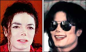
Picture 4. Patient in 1993 and 1996. Post-vitiligo universalis, and subsequent bleaching treatment. Photos taken from BBC News online.
Immuno Suppressive Treatments:
As vitiligo is believed to be an autoimmune disorder, supressing the immune system would then be an effective treatment at halting the spread of vitiligo and even inducing repigmentation. Corticosteroids are the main choice for this treatment and they effectively halt the progressive spread of vitiligo and also lead to repigmentation of lesioned areas. For small, localized lesions, topical and intralesional corticosteroids are used once a day. For rapidly spreading vitiligo or vitiligo universalis, systemic corticosteroids are employed; however, the role of systemic corticosteroids is controversial due to the serious adverse side effects.
Non-surgical Treatment: phototherapy and photochemotherapy
Currently, the most popular treatments for vitiligo are forms of phototherapy; it is known that various sources of UV light can be successfully used to stimulate repigmentation. Phototherapy can either be used by itself or in conjunction with light-sensitizing drugs.
PUVA treatment is the most popular treatment for vitiligo currently; the concept for the treatment dates back thousands of years, when the plants Psoralea coryifolia Linnaues and Ammi majus Linnaeus where eaten or used topically in Egypt and India to treat vitiligo. Today, isolates of the plants are used topically or orally in conjunction with a synthetic compound to chemically increase sensitivity to light. The patient is then exposed to a measured amount of natural sunlight (PUVASOL) or artificial UV radiation (PUVA) to induce re-pigmentation.
The amino phenylalanine is also used to treat vitiligo, although it is not a photo-sensitizing chemical. It is known that phenylalanine is a precursor for melanin via L-tyrosine, and it appears that there is a problem with L-phenylalanine metabolism in vitiligo. A combination of topical application and oral ingestion of phenylalanine with natural sunlight exposure resulted in repigmentation for 83% of patients.
In addition to photochemotherapy, there are several forms of phototherapy, without the light-sensitizing chemicals.
UV light in the wavelengths from 290-320 nm are often used to treat vitiligo. There are few studies reporting the efficacy of this treatment, but anecdotally the treatment appears effective, though not as good as other forms of phototherapy. In addition to broad band UV (BUVB), there is also narrow band UV treatment (NBUVB). This is a more recent form of UV phototherapy and was initially used to treat psoriasis. This UV treatment operates between 305 and 311 nm, and is highly effective for treating psoriasis and moderately effective at treating vitiligo. When used as monotherapy, NBUVB is slightly but not significantly more effective at stimulating repigmentation than PUVA treatment.
In addition to UV treatment, the excimer laser has now been implemented as phototherapy for vitiligo. The 308 nm laser produces light similar to the narrow band UVB treatment, and has shown good results in stimulating repigmentation of about 90% of patients. The excimer laser also allows more focused treatment on specific lesions, or hard to reach areas of the body.
There is little scientific evidence examining why phototherapies are effective at stimulating repigmentation; however, it is very strongly believed that if there are stores of unaffected but immature melanyocytes in the lesioned area, these melanocytes can be induced to produce melanin in the lesioned areas. Likewise, there are often functional, but not melanin producing melanocytes at lesion boundaries that can produce melanin that spreads into the lesions causing repigmentation.
Surgical Treatment:
In addition to phototherapy, there is also surgical treament for vitiligo that mainly consists of grafting patches of skin with healthy melanocytes to lesioned sites. In both minigrafting and suction blister grafting, superficial layers of skin are removed from normally-pigmented skin and these patches then grafted onto the lesioned areas. These techniques are believed to work by healthy melanocytes from the grafts proliferating into the lesioned areas and repopulating them. Success rates for these techniques are between 80% and 90%. Full thickness grafts of skin are implanted in minigrafting, using 1-2 mm healthy skin punches from areas such as the buttocks and followed by exposure to sunlight or PUVA treatment. Success of minigrafting is around 67% in patients.
The future of surgical treatment for vitiligo is actually extracting melanocytes from a donor area of the patient's healthy skin, culturing these melanocytes into a large population, then grafting them as sheets onto the lesioned areas. Preleminary trials with this technique have been highly successful; however there is some concern about controlling malignancy in the cultured melanocytes.
Bleaching Treatment:
In addition to treatments to repigment skin, in extremely progressive,
full-body vitiligo called vitiligo universalis, the patient can opt to bleach
the remaining pigment off healthy skin. This treatment does have profound psychosocial
implications, especially for naturally dark-skinned individuals who become light
or white-skinned. This is the proposed medical scenario with performer Michael
Jackson, who claims to have been afflicted with vitiligo universalis. As opposed
to seeking repigmentation, Jackson opted instead for depigmentation of remaining,
naturally dark areas. This is a feasible scenario, although there is great debate
as to the authenticity of Jackson's claim (Taneja 2002).
References
Janeway, C. A. Jr., Travers, P., Walport, M., and Shlomchik, M. J. Immunobiology 5. New York: Garland Publishing, 2001.
Huang, C.L., Nordlund, J.J., & Boissy, R. 2002. "Vitiligo: A manifestation of apoptosis?" American Journal of Clinical Dermatology 3(5): 301-308.
Majumder, P.P., Das, S.K., Li, C.C. 1988. "A genetical model for vitiligo." American Journal Human Genetics 43: 119-125.
Majumder, P.P., Nordlund, J.J., Nath, S.K. 1993. "Pattern of familial aggregation of vitiligo." Archives of Dermatology 129: 994-998.
Nath, S.K., Majumder, P.P., Nordlund, J.J. 1994. "Genetic epidemiology of vitiligo: multilocus recessivity cross-validated." American Journal of Human Genetics 55: 981-990.
Njoo, M.D., & Westerhof, W. 2001. "Vitiligo: Pathogenesis and Treatment." American Journal of Clnical Dermatology 2(3): 167-181.
Prota, G. 2000. "Melanins, Melanogenesis and Melanocytes: Looking at Their functional significance from the chemist's viewpoint." Pigment Cell Research 13: 283-293.
Taneja, A. 2002. "Treatment of Vitiligo." Journal of Dermatological Treatment 13: 19-258.
Davidson College Biology Bio 307
Spring Semester 2003
Contact Peter Leese or View Immunology at Davidson College