This web page was produced as an assignment for an
undergraduate course at Davidson College.
Terminal Deoxynucleotidyl Transferase (TdT)
-
Introduction
-
Overview of Development of Lymphocyte
Diversity
-
Normal Function of TdT
-
Effect of Missing TdT
-
Drug Interactions with TdT Activity
-
Laboratory Applications of TdT
-
References
Introduction
The transfer of genetic information from one generation
to the next is dependent on accurate replication of parental DNA.
To accomplish this goal, DNA polymerases (along with many other enzymes)
have evolved which are able to copy DNA from a template strand very effectively.
However, not all DNA polymerization that takes place in an organism is
based on a template. Terminal deoxynucleotidyl transferase (TdT)
is a template-independent DNA polymerase which is capable of catalyzing
the elongation of a DNA strand by the addition of nucleotides from the
surrounding solution. Such nucleotide addition is required in a few
specific situations, the most studied of which is lymphocyte development.
This web page will explore the role of TdT in the development of lymphocyte
diversity.
Return to the Top of the Page
Overview of Development
of Lymphocyte Diversity
Adults have an incredibly diverse repertoire of B and
T cell antigen receptors. It has been estimated that the numbers
approach 1014 different specific immunoglobins
(Ig's) and 1018 distinct T cell receptors
(TCR's). This incredible diversity greatly exceeds the number of
different genes in the entire human genome, making it impossible for each
specific antigen receptor to be coded by an individual gene. Instead,
mechanisms have evolved which derive a large amount of diversity from a
relatively small number of genes. This web page will focus on the
development of diversity in Ig's, but it should be noted that similar mechanisms
are at work in the development of diversity in TCR's. In humans,
there are several mechanisms which work together to generate Ig diversity.
Ig's are composed of two identical light chains combined with two identical
heavy chains, all four of which are composed of a variable (V) region and
a constant (C) region. Since each individual can form many different
light and heavy chains, association of different light and heavy chains
leads to significant diversity. Further diversity is obtained in
the formation of individual light and heavy chain V-regions by splicing
together different regions of the gene segment to form an active exon.
These regions, called V and J in the light chain and V, D, and J in the
heavy chain, are present in multiple copies in each gene segment.
Therefore the choice of specific V(D)J joining adds considerable diversity
to the immunoglobin. The amount of diversity is increased yet again
by the fact that the splicing of V, D, and J regions described above is
not precise. Nucleotides are often added to or deleted from the joint.
Nucleotides can be added to the joint by two methods. The first method
occurs through the formation and cleavage of hairpins during DNA splicing
and results in the insertion of pallindromic segments of DNA (P-nucleotides).
The second method is the random insertion of non template (N) nucleotides,
which is catalyzed by TdT. Diversity is increased yet again by a
process known as somatic hypermutation much later in the process of immunoglobin
development (Janeway et al., 1999). This page will focus on
the role of TdT in the insertion of N-nucleotides. For a more detailed
look at the development of lymphocyte diversity, check out Dr. Campbell's
somatic
recombination animation (Campbell, 2000).
Return to the Top of the Page
Normal Function of TdT
Background
Early research in to the joining of D and J segments led
to the discovery that some nucleotides are present in the joint that were
not part of either segment before they were joined (Alt et al.,
1982). This study was among the first to implicate TdT as a possible
mechanism for the insertion of these nucleotides. This conclusion was based
on TdT's high concentration in the thymus and bone marrow (where such recombination
events occur) and the fact that many of the inserted nucleotides were guanine
(G) residues, which TdT had been shown to prefer over other nucleotides
(Alt et al., 1982). Later research has shown that in addition
to its G utilization preference, TdT is affected by steric interactions
of nucleotide stacking which favor the insertion long strings of purines
or pyrmidines, making the process somewhat less random than previously
thought (Gauss et al., 1996). The rate of nontemplated DNA
addition by TdT has been found to be at least 76-fold slower than that
of templated synthesis (Clark, 1988).
Regulation of Expression
Because TdT is capable of adding non template nucleotides
to any exposed 3' end of DNA, it is very important that the body limits
its expression to times and places where its function is beneficial.
Studies in mice have shown that TdT is not detected in neonatal thymi,
but begins to appear about four days after birth (Bogue et al.,
1993). There is then a one to two day lag period before N-nucleotide
addition is observed (Bogue et al., 1992). It has been suggested
that the lack of neonatal TdT is related to the smaller amount of self-reactivity
observed in immunoglobins produced without the addition of N-nucleotides
(Bogue et al., 1992). If one considers that a fetus is still
under the immune protection of the mother, it makes sense that the added
immunoglobin diversity that N-nucleotide addition provides is not selected
for strongly enough to outweigh the problem of creating many self-reactive
antibodies.
Expression of TdT is tightly controlled in adults as well.
TdT expression in the thymus and bone marrow closely follow that of RAG-1
and RAG-2, which combine to form a complex which cuts one strand of the
DNA prior to V(D)J joining. The only difference in expression is
that TdT is expressed only during stages while heavy chain rearrangement
is occurring while RAG-1 and RAG-2 are expressed during both heavy and
light chain rearrangement (Li et al., 1993). This tight control
of expression leads to the observation that N-nucleotide addition is common
in heavy chain rearrangement and very rare in light chain rearrangement.
The stage in B cell development in which the heavy chain is rearranged
is known as the pro-B cell stage. Therefore only cells in this stage
exhibit TdT. Staining for TdT can reveal the location of these cells
as shown in Figure 1 below.
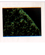
Figure 1: Part of a transverse section of rat femur showing
fluorecence labeled TdT cells (green). These cells are most highly
concentrated toward the outside of the bone (upper right corner) and less
numerous toward the middle of the bone (lower left corner) because more
developed cells that no longer express TdT are moving toward the middle
of the bone. (Photograph courtesy of D. Opstelten and M. Hermans.
From Janeway et al.,Immunobiology, 4th ed., Garland Publishing,1999,
p. 198)
Recent studies have shown that regulation of TdT
expression is controlled on the DNA level by the selective methylation
of promoter regions of genes which code for the protein (Nourrit et al.,
1999). It has also been shown that the RAG-1/RAG-2 complex plays
a vital role in the regulation of N-nucleotide addition by persisting on
the DNA after single strand cleavage, blocking TdT from adding nucleotides
until both strands are cleaved (Grawunder et al., 1997).
Although the average number of N-nucleotides added to
each junction correlates with the amount of TdT expression, it is thus
far impossible to predict the number of N-nucleotides which will be added
to a given junction. It has been shown that the level of diversity
is not coded in the stem cell and is probably dependent on environmental
factors (Bogue et al., 1993). Early work predicted that N-nucleotides
would be added until a homology of one nucleotide occurred, at which point
templated DNA polymerases would finish the joint (Alt et al., 1982).
However, later work has shown that such homologies are not necessary for
N-nucleotide addition to stop, nor does their presence dictate a stop in
N-nucleotide addition. Junction formation has been observed in joints
in which zero, one, two, or more homologies exist (Komori et al.,
1993).
Return to the Top of the Page
Effect of Missing TdT
Studies on mice have shown that animals which lack TdT
expression have en extremely small number of N-nucleotides (Gilfillan et
al.,
1993). This lack of N-nucleotides limits the number of different
antigen receptors seen in these mice due to fewer possible recombinations.
However, the observed decrease in diversity was much greater than could
be explained by this logic alone. Apparently, recombination in mice
is much more strongly governed by homology than it is in humans, leading
to the preferential formation of certain junction combinations at the expense
of others. TdT driven addition of N-nucleotides eliminates this bias
for certain pairings and therefore plays a more important role than expected
in the generation of diversity in mice (Gilfillan et al., 1993).
Return to the Top of the Page
Drug Interactions with TdT Activity
TdT adds nontemplate nucleotides to DNA sequences with
an exposed 3' end. This addition depends on the presence of a hydroxyl
group at the 3' position on the terminal nucleotide. Therefore, nucleotide
addition can be limited to one nucleotide if a modified nucleotide lacking
a 3' hydroxyl group is present in the solution. One such nucleotide,
[alpha]cordycepin-5'-triphosphate, has been shown to accomplish this limitation
(Promega, 2000). This interaction may prove applicable to the treatment
of refractory TdT-positive leukemia, a type of leukemia in which the cancerous
cells are TdT+. One such treatment
involving the nucleotide described above is currently in phase I of study
(Division of Hematology / Oncology at New England Medical Center, 1999).
Return to the Top of the Page
Laboratory Applications of TdT
Because of its ability to add nucleotides to any DNA strand
with a free 3' end, TdT has been adopted as a useful tool in immunology
research. When cells undergo apoptosis, their DNA is cleaved into
many small segments which diplay free 3' ends. These apoptotic cells
can be labeled with biotin-coupled uridine by exposing them to a solution
containing TdT and these uridine residues. These biotin labels can
then be detected with enzyme bound to streptavidin, which is known to bind
to biotin. If the subtrate to this ezyme, a colorless solution, is
added to the culture, a colored precipitate will be produced in cells which
have undergone apoptosis (Janeway et al., 1999). A similar
method which uses fluorescence techniques instead of biotin labeling has
also been developed to locate apoptotic cells (Mitra, 1996).
Return to the Top of the Page
References
Alt, F.W. and D. Baltimore. 1982. Procedings
of the National Academy of Science, USA. 79, 4118-4122.
Bogue, M., S. Gilfillan, C. Benoist, and D. Mathis. 1992.
Procedings
of the National Academy of Science, USA. 89,11011-11015.
Bogue, M., H. Mossmann, U. Stauffer, C. Benoist, and D.
Mathis. 1993. European Journal of Immunology.
23,
1185-1188.
Campbell, A.M. 2000. Flash Animation of Somatic
Recombination of Antibody Light Chain. <http://bio.davidson.edu/Biology/Courses/Immunology/Flash/somaticrecomb.html>
Accessed 2000 March 1.
Clark, J.M. 1988. Nucleic Acids
Research. 16, 9677-9686.
Division of Hematology / Oncology at New England Medical
Center. 1999. Cancer Clinical Trials Office: Cordycepin.
<http://www.nemc.org/hemonc/trials/cordycep.htm>
Accessed 2000 Feb 28.
Gauss, G.H. and M.R. Lieber. 1995. Molecular
and Cellular Biology. 16, 258-269.
Gilfillan, S., A. Dierich, M. Lemeur, C. Benoist, and
D. Mathis. 1993. Science. 261, 1175-1178.
Grawunder, U. and M.R. Lieber. 1997. Nucleic
Acids Research. 25, 1375-1382.
Janeway, C.A., P. Travers, M. Walport, and J.D. Capra.
1999.
Immunobiology, 4th ed. Garland Publishing, New York.
Komori, T., A. Okada, V. Stewart, and F.W. Alt. 1993.
Science.
261, 1171-1175.
Lewis, S.M. 1994. Advances in Immunology.
56,
27-150.
Li, Y.S., K. Hayakawa, and R.R. Hardy. 1993.
Journal
of Exp. Medicine. 178, 195-960.
Mitra, G. 1996. Detection of Apoptotic Cells
using the Apoptosis Detection System, Fluorescein: Promega Notes
Magazine (57). <http://www.promega.com/pnotes/57/5573b/5573b.html>
Accessed 2000 Feb 28.
Nourrit, F., I. Coquilleau, M.F. D'Andon, F. Rougeon,
and N. Doyen. 1999. Journal of Molecular Biology.
292,
217-227.
Promega Corporation. Feb 2000. Abstract For
Terminal Deoxynucleotidyl Transferase. <http://www.promega.com/tbs/tb088/tb088.html>
Accessed 2000 Feb 28.
Return to the Top of the Page
Return to Bob Magnussen's Immunology
Home Page
Return
to Immunology Main Page

Send comments, questions, and suggestions to: bomagnussen@davidson.edu