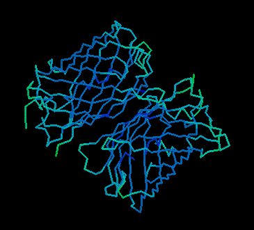
fig 1. A still image from gfpdimer.pdb. (GFP, though a monomer in vivo, crystalizes as a dimer.) Click to view (requires Rasmol).
Green Fluorescent Protein as a Reporter Gene
By Ben Buxton
"If you don't already use GFP, you will. Maybe not today, maybe not tomorrow, but soon... and for the rest of your life"
Green-fluorescent protein (GFP), one of the hottest new biological tools, is responsible for the stunning bioluminescence of the Pacific Northwest Jellyfish, Aequorea victoria. While in its native host, GFP emits a green colored light when it accepts blue light from a calcium activated photoprotein. Given another source of blue light, such as UV, which cannot be detected by human eyse, GFP will produce a green light. A human using a UV light will therefore only see GFP (and other fluorescents), because it will be the only source of visible light. It is just this property that makes the clone of the GFP gene a powerful tool for use in the lab. Since the cloning of the GFP gene in 1992 by Douglas Prasher (of USDA/APHIS) it has been demonstrated that the GFP gene can be expressed in non-homologous species, from yeast and plant cells to Drosophila and vertebrates, including humans1. It seems that this lack of specificity to jellyfish is because GFP requires no substrates (other than oxygen) to fluoresce, and has no physiological effect on cell operation. In a sense a transformed cell doesnÕt ÒknowÓ that GFP is even there, and therefore can remain alive while being studied.
Such a powerful tool has many applications in biological research and commercial biotechnology including: Òmeasuring gene expression in vitro, selecting transgenic cells, studying fusion proteins, studying intracellular protein traffic (and thus identifying signal sequences), determining cell lineage, assessing promoter activity, developing cell- and tissue-specific markers, investigating the pathogen movement and disease development, biomonitoring of organisms released into the environment, developing bioindicators for detecting environmental pollutants, ensuring the containment of genetically modified organisms and in evolutionary and ecological studies of transgenic organisms (Prasher 1995 Trends in Genetics 11: 320-323).Ó1
The main concern of this paper, however, is with the application of GFP as a reporter gene. Reporter genes are common biological tools that demonstrate clearly when they a gene is turned on or off. For instance, the Lac-Z gene (coding for Beta-Galactosidase), when uninterrupted and functional, will change the colorless substrate X-gal to blue. If functional Lac-Z is part of a plasmid in a bacteria or yeast colony, it will change the cells from from white to blue. This so called blue white selection is commonly used to find cells with recombinant DNA or plasmids (cells with an insert interrupting Lac-Z), after performing a transformation (introducing new DNA into a cell). GFPÕs distinctive properties allow it also to be used as a reporter of gene expression.
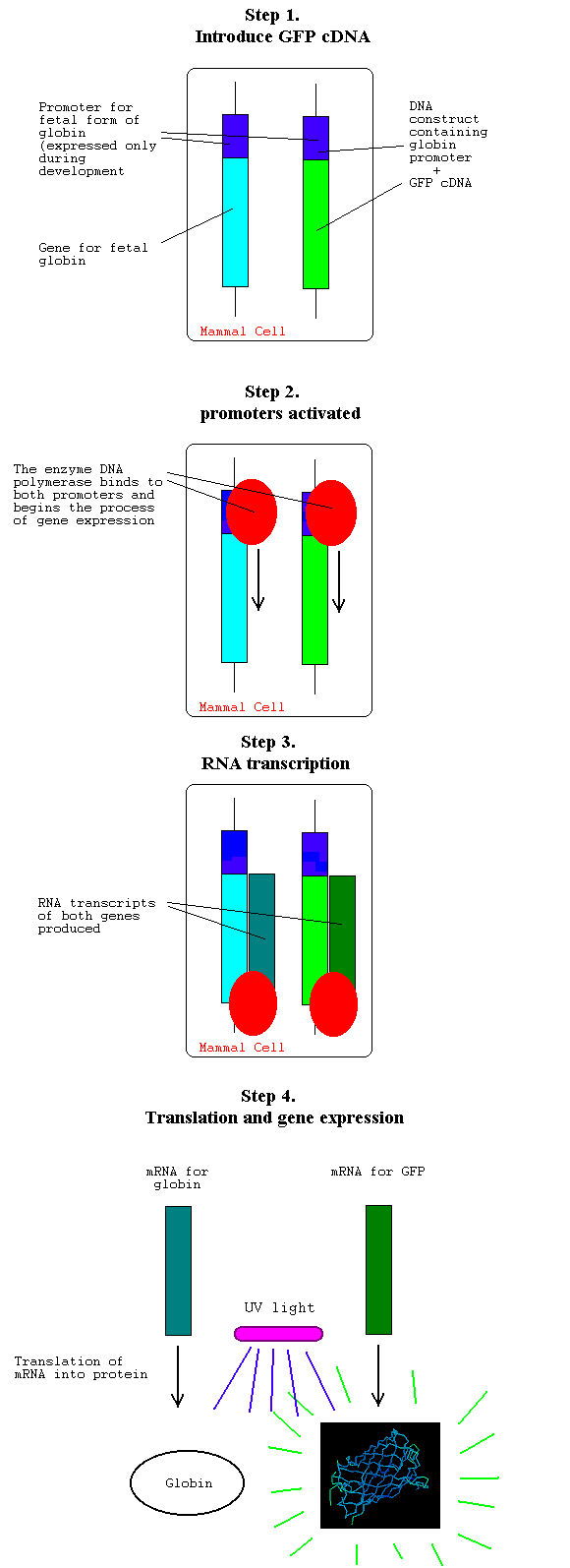
Natures method of gene expression, where the proteins that make up a cell are made from their DNA codes, gives molecular biologists a useful tool. Each gene in an organism is located after the promoter and inducer sequences specific to it. These are portions of DNA occurring before the actual coding sequences that an RNA polymerase enzyme binds to in order to begin translation of the gene. In a little understood process, certain promoters have a property which makes them bind more often to RNA polymerase then other promoters, and thus proteins are expressed in different amounts. In other words, the cell does not really ÒknowÓ what genes it is translating, it only ÒknowsÓ which promoters are being used. Since the cell only sees the promoter, the gene for GFP can be attached downstream from the promoter (and inducer) DNA sequences for whatever gene one wishes to study. If this DNA construct is introduced into a cell, whenever the gene under study is expressed in the cell, the GFP produced simultaneously will fluoresce green under a UV lamp. Rather than having to detect the protein of interest, say with antibodies, or by various other destructive methods you can watch the living, normally functioning cell and clearly determine when the gene is turned on and off.
The following series of figures illustrate how GFP could be used to "report" when a gene encoding a fetal form of globin, a hemoglogin subunit, is being expressed: (figure by Ben Buxton)
Using GFP as a reporter gene is not without its potential problems. A major one is that only one gene can be tracked at a time. To solve this problem, reserachers are mutating the chromophore, or light emitting portion of the protein, in attempts to find a mutant form of the gene that would fluoresce in a different color. In addition, GFP signals can be weak and hard to detect without high expression. Some cells will naturally fluoresce, too, such as lignin in plant cells. This could make bona fide GFP fluorescence hard to detect.
Citations:
1Prakesh, C.S. 1997 Sep. Green Fluorescent protein. <http://199.88.21.38/AgHe%20Website/Biotech%20Website/Prakash_Publications%7f%7f%7f%7f%7f/green_protein.html> Accessed 1998 Feb 18.
![]()
![]()
Direct correspondence to: bebuxton@davidson.edu