This web page was produced as an assignment for an undergraduate course at Davidson College
My Favorite Protein/Gene: Polycystins
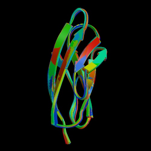
Figure 1. This is a depiction of domain 1 for the protein polycystin-1. It consists of 80 of the 4303 amino acids in the entire protein. (Structure Explorer- Polycysin-1).
The purpose of this website is to describe the function and structure of Polycystin-1 and -2, which are vital proteins to the development and maintenance of the kidneys.
Polycystins
Polycystins are proteins that have been found to play important roles in cell
to cell and cell to extracellular matrix interactions (Gallagher, 2002). Research
into the function of these proteins has also revealed that they play a part
in cilia action in the nephrons and calcium channeling. Boletta et al. have
reported that polycystin-1 control renal tubular differentiation, cell proliferation,
and apoptosis (2000). The coordination of these functions adds up to the maintenance
and regulation of renal epithelial cell growth. It has also been found that
polycystin-1 may have a role in the structural integrity of blood vessels (Kim,
et al., 2000).
Polycystin-1
Polycystin-1 is an integral membrane protein that is the translational product
of the PKD1 gene found on chromosome 16 (OMIM Polycystic-1, 2003). At 4303 amino
acids long (NCBI-1, 2002), it spans the plasma membrane 11 times with a ~3000
amino acid extracellular domain and a small cytoplasmic tail. Polycystin-1 is
found in the basolateral membrane and complexed to polycystin-2 (Polycystin-2
Projects, 2003). This protein is expressed in the epithelial cells of the kidneys
as well as that of the vasculature (Kim). In the kidneys, polycysin-1 is localized
to the cilia (Ong, 2003), (Surya et al., 2003). Despite all the research that
has illuminated the structure and function of polycystin-1, the mechanism by
which it works has yet to be discovered.
Polycystin-2
Polycystin-2 is also a integral membrane protein, but it is found on chromosome
4 (OMIM). It is located mostly within the endoplasmic reticulum membrane (Polycystin-2
Projects). It is 968 amino acids long (NCBI-2, 2002). There are two different
models for where and how this protein functions, as shown in Figure 2.
Figure 2. Current debate on the site of polycystin-2 (PC2) channel action. PC2
channels on both the endoplasmic reticulum and the cell membrane are shown.
PC2 channels can be translocated to the cell membrane in the presence of PC1
or defined chemical chaperones and proteasome modulators. It is possible that
the PC2 channel might function on either membrane in a cell-specific manner.
Abbreviations: IP3R, inositol (1,4,5)-trisphosphate receptor; SERCA, sarcoendoplasmic
reticulum Ca2+-ATPase. (Stayner, 2001) permission pending.
Wherever this protein actually resides, it is in close association with polycystin-1 and the plasma membrane. The main function of this protein is thought to be a calcium channel. Polycystin-2 is thought to function as a “calcium-regulated, calcium-permeable cation channel” that is important for the formation of kidney tubules (Ikeda et al., 2002). This form of the protein has only six membrane spanning domains. The sequence of this protein is similar to the last six membrane spanning domains of polycystin-1 (Polycystin-2 Projects).
Polycystin-1 and –2 are connected by homotypic interactions between their two C-terminal cytoplasmic tails. Their interaction is necessary for the normal tubulogenesis and renal tubule function(OMIM). See Figure 3.
Figure 3. Polycystin (PC) domain architecture. Only PC1 and PC2 are shown. An
11-transmembrane topology is shown for PC1, with respective domains indicated.
The inset figure shows a polycystic kidney from a patient with autosomal dominant
polycystic kidney disease (ADPKD). Abbreviations: EF-hand, helix–loop–helix;
GPS, G-protein-coupled receptor proteolytic site; LDL, low-density lipoprotein;
LH2, lypoxygenase homology 2; P, pore region; PKD, polycystic kidney disease;
REJ, sperm receptor for egg jelly, sea urchin homolog; WSC, cell wall integrity
and stress response component. (Stayner, 2001) permission pending.
Orthologs in other Species
Several orthologs have been found in other animal species such as mice and canines.
Pdk1, as it is referred to in these animals, produces as similar protein, polycystin-1
in both of these species. The canine ortholog has been found to be more similar
to the human gene than the mouse gene is to the human one. This has led researchers
to use the Madin Darby canine kidney (MDCK) cell line for research on the role
of polycystin-1 in cystogenesis and tubulogenesis (Dackowski, 2002).
The study of the ortholog LOV-1 in Caenorhabditis elegans has also been helpful
in elucidating the function of polycystin-1 in the morphological development
of the kidney tubules (Kaletta et al., 2003).
Mutation leads to Polycystic Kidney Disease (PKD)
Mutations in either polycystin-1 or –2 account for the majority of Autosomal
dominant PKD. This disease, which is characterized by abnormal growth of cysts
in the kidneys as well as the liverand other organs, affects about 500,000 people
in the United States. (Polycystic Kidney Disease, 2002).
The cysts that grow in the kidney are the result of any number of mutations
within the genes PKD1 and PKD2 that code for polycysin-1 and –2, respectively.
Over 90 mutations have been discovered in PKD1 alone, which lead to the disruption
of proper function. These mutations have been found to be missense, nonsense,
and frameshift. And as mentioned above, proper interaction between polycystin-1
and –2 is required for the complex to work so a mutation in one causes
a malfunction in both proteins (Eo, 2002).
Reeders (1992) proposed a two-hit model for mutations in PKD1. His hypothesis
was that PKD results somatic and/or germline mutations of both copies of PKD1.
This is illustrated in Figure 4.
Figure 4. 'Two-hit' model of cystogenesis in autosomal dominant polycystic kidney
disease (ADPKD). (a) All the epithelial cells in a renal tubule contained the
same germline PKD mutation. (b) Somatic mutation of the 'wild-type' PKD allele
occurred within an individual epithelial cell. (c,d) Clonal proliferation of
this epithelial cell results in tubular dilatation and cyst formation. (York,
2001) permission pending.
Conclusion
There is still much research that must be done in order to fully understand the important role that polycystins play in kidney development. Certainly, studying the effects of mutated proteins and the effects they have on tubulogenesis versus cystogenesis is a start in the right direction. The many roles that polycystins have in kidney function necessitate that a research continues in this field.
References
Boletta, A. et al. 2000. Polycystin-1, the Gene Product of PKD1, Induces Resistance to Apoptosis and Spontaneous Tubulogenesis inMDCK Cells. Molecular Cell. 6:1267-1273.
Dackowski, W.R. et al. 2002 Jul. Canine PKD1 is a single-copy gene: genomic organization and comparative analysis. Genomics. 80(1):105-12.
Eo, H.S. et al. 2002 Aug. Three novel mutations of the PKD1 gene in Korean patients with autosomal dominant polycystic kidney disease. Clinical Genetics. 62(2):169-74.
Gallagher, A.R. et al. 2002 Apr. Molecular basis of autosomal-dominant polycystic kidney disease. Cell Molecular Biology Science. 59(4):682-93.
Ikeda, M. and W.B, Guggino. 2002 Sep. Do polycystins function as cation channels? Current Opinion in Nephrology and Hypertension. 11(5):539-45.
Kim, K. et al. 2000 Feb 15. Polycystin 1 Is Required for the Structural Integrity of Blood Vessels. Proceedings of the National Academy of Sciences of the United States of America. 97(4): 1731-1736.
Kaletta, T. et al. 2003 Jan. Towards understanding the polycystins. Nephron Experimental Nephrology.93:e9-e17
NCBI Sequence Viewer- Polycystin-1. 2002 August 5. <http://www.ncbi.nlm.nih.gov/ entrez/query.fcgi?cmd=Retrieve&db=protein&list_uids=1730587&dopt=GenPept> Accessed 2003 Mar 13.
NCBI Sequence Viewer- Polycystin-2. 2002 Aug 5. <http://www.ncbi.nlm.nih.gov/ entrez/query.fcgi?cmd=Retrieve&db=protein&list_uids=6174909&dopt=GenPept> Accessed 2003 Mar 13.
OMIM- Polycystic Kidney Disease 1. 2003. <http://www.ncbi.nlm.nih.gov/ entrez/dispomim.cgi?id=601313> Accessed 2003 Mar 13.
Polycystic Kidney Disease. 2002 May. National Kidney and Urologic Diseases Information Clearinghouse. <http://www.niddk.nih.gov/health/kidney/pubs/polycyst/ polycyst.htm> Accessed 2003 Mar 13.
Polycystin-2 Projects.<http://info.med.yale.edu/pharm/hermen/polycystinprojects.html>
Accessed 2003 Mar 13.
Ong, A.C. and D.N. Wheatley. 2003 Mar 1. Polycystic kidney disease-the ciliary
connection. Lancet . 361(9359):774-6.
Reeders, S.T. 1992 Jul. Multilocus polycystic disease. National Genetics. 1(4):235-7.
Stayner, C. and J. Zhou. 2001. Polycystin channels and kidney disease
Trends in Pharmacological Sciences. 22:543-546. <http://reviews.bmn.com/rsearch/section/record?uid=TIPS.etd00974_01656147_v0022i11_00001832>
Accessed 2003 Mar 13.
Structural Explorer- 1B4R. <http://www.rcsb.org/pdb/cgi/explore.cgi?job= graphics;pdbId=1B4R;page=;pid=262631047629510&opt=show&size=500> Accessed 2003 Mar 13.
Surya, M.N. et al. 2003 Jan 6. Polycystins 1 and 2 mediate mechanosensation in the primary cilium of kidney cells. Nature Genetics. 33(2): 129-137.
York, P. 2001. A 'two-hit' model of cystogenesis in autosomal dominant polycystic kidney disease? Trends in Molecular Medicine. 7:151-156. <http://reviews.bmn.com/rsearch/section/record?uid=MMT.etd00704_14714914_v0007i04_00001953> Accessed 2003 Mar 13.
For questions or comments, email Heather Maloney