This page was created as an undergraduate assignment for a molecular biology course.


Molecular Method: Fluorography

Presented by Isaac Miller


I. Introduction to fluorography:

Fluorography is a method used to visualize substances present in gels, blots, or other biochemical separations. In fluorography, radioactively labeled substances emit radiation that excites a molecule known as a fluor or scintillator that is present in the gel. When the excited molecule relaxes to its ground state, it emits a photon of visible or ultraviolet light that is detected by photographic film. The developed film indicates which bands in the gel (or spots on the TLC, etc.) contain radioactively labeled material (Waterborg et al., 1994).


II. Radioisotopes

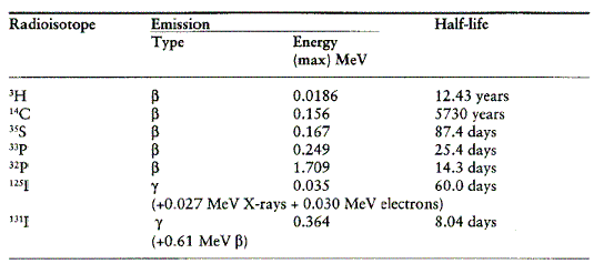
Radioisotopes are atoms with unstable nuclei that decay over time, emitting detectable radiation. In biology, radioisotopes are often incorporated into molecules of interest so that those molecules can be followed by following their radiation trail. The table below lists radioisotopes used often in biology with their corresponding half-lives and the energies of their emitted radiation (Laskey, 2003).

Table 1. Half-lives and radiation energies of radioisotopes used often in biology ( Laskey, 2003). Click on the table to view the document from which it was obtained in Adobe Acrobat PDF format. Permission for use of this table is pending.
III. Purpose of fluorography

Directly detecting the radioactive signals emitted by the labeled substances is known as autoradiography, but sometimes the signal is too weak to expose the photographic film or is absorbed by the gel before it can reach the film. In these cases, a technique to amplify weak radiation signals is needed. The major benefit of fluorography over autoradiography is that it offers increased sensitivity to weak radiation signals. Fluorography allows a greater than ten-fold increase in sensitivity for samples labeled with 35S and an approximately 1000-fold increase in sensitivity for samples labeled with 3H (Laskey, 2003).


IV. General mechanism

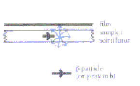
Radiation from ß-emitting isotope-labeled molecules excites molecules of a fluor, such as 2,5-diphenyloxazole (PPO) (Prahl, 2001) or Amersham Biosciences' reagent Amplify™, that has been added to the gel. The excited fluor molecules fluoresce; that is, they emit visible light as they quickly return to their ground state through allowed electronic transitions. The visible light emitted by the fluor molecules is detected by photographic film, rendering the fluorograph. This general mechanism is outlined in the figure below (Laskey, 2003).

Figure 1. Overview of fluorography: a ß-particle with a short lifetime is emitted by the labeled substance being followed. The ß-particle excites a molecule called a fluor or scintillator, which subsequently emits a photon of visible or ultraviolet light, which passes through the gel and is detected by film. Permission for use of this image is pending. To access the document in PDF format from which this figure is taken, click on the image.


V. Structure of a common fluor: PPO

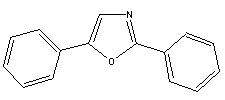
Figure 2. Structure of PPO (from www.chemfinder.com)

Radioactive energy from the molecule to be detected promotes an electron from the highly conjugated PPO into a higher energy level. As this electron returns to the ground state energy level, a photon of light is emitted. Thus, the presence of the original radioactive signal is indirectly detected (Carroll, 1998).


VI. Useful link:

For additional information concerning the theory behind fluorography or for method protocols, see this pamphlet regarding autoradiography, fluorography and chemiluminescence from Amersham Biosciences or see Waterborg, et al.


VII. References

Carroll FA. 1998. Perspectives on Structure and Mechanism in Organic Chemistry. New York: Brooks/Cole Publishing Company. p 811-814.

Laskey RA. 2003. Reveiew 23: Efficient Detection of Biomolecules by Autoradiography, Fluorography or Chemiluminescence. Amersham Biosciences homepage. <http://www4.amershambiosciences.com/aptrix/upp00919.nsf/(FileDownload)?OpenAgent&docid=1DC784945E4EABB3C1256B26000DCFDB&file=review23_part1.pdf> Accessed 2003 Feb 18.

Prahl S. 2001. 2,5-Diphenyloxazole (PPO). Oregon Medical Laser Center homepage. <http://omlc.ogi.edu/spectra/PhotochemCAD/html/PPO.html> Accessed 2003 Feb 18.

Waterborg JH, Matthews HR. 1994. Fluorography of Polyacrylamide Gels Containing Tritium. In: Walker JM, editor. Basic Protein and Peptide Protocols. v 32. Totowa, New Jersey: Humana Press. p 163-167.


Molecular Biology Homepage

Return to my Web Page List

Send questions or comments to Isaac Miller

Last modified: Feb. 18, 2003