*This web page was produced as an assignment for an undergraduate course at Davidson College.*
What is FISH?
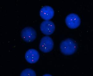
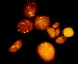
These images were borrowed from The University of Texas MD Anderson Cancer Center web page and can be accessed at http://www3.mdanderson.org/depts/pathology/fish/fish.html. Permission for the use of these images is pending.
Fluorescence In-Situ Hybridization (FISH) is a simple and fast research tool that utilizes fluorescence techniques for metaphase analysis of chromosomes. According to Nikon Microscopy, in-situ hybridization has been around for a quarter of a century as a method of researching the DNA sequences of chromosomes and cells (Nikon Microscopy). Only in the last 15 years have fluorescence techniques been utilized (Nikon Microscopy). FISH uses these fluorescence techniques to label genes and chromosomes, which is useful for gene mapping and detecting chromosomal abnormalities (click to find out details about how FISH works).
Davidson College Molecular Biology Homepage
Send questions or comments to brstonestreet@davidson.edu