My Favorite Yeast Genes:
PDI1 is an annotated gene located on Saccharomyces cerevisiae chromosome 3. PDI1 encodes a protein disulfide isomerase, present in the endoplasmic reticulum, which plays an essential role in the formation of disulfide bonds in secretory and cell-surface proteins (Dolinski 2004). While the function of PDI1 is well-known, the neighboring YCL047C hypothetical ORF is entirely uncharacterized. This ORF’s cellular component, molecular function, and biological process are still unknown (Dolinski 2004).
My Favorite Annotated Yeast Gene: PDI1
Chromosomal Location
PDI1 is located within a small region of Saccharomyces cerevisiae chromosome 3, as shown below.

Biological Process
Molecular Function
The PDI1 protein contains two thioredoxin-like domains (see figure 2), each with a copy of the active site sequence motif CGHC (Norgaard 2001).

Figure 2 Thioredoxin-like domains of PDI1. This image depicts the relative position and extent of the thioredoxin-like domains (open boxes) within the PDI1 protein. CGHC active site sequence motifs, which form an intramolecular disulfide bond capable of converting a pair of sulfhydryl groups in a polypeptide substrate into a disulfide bond, are also shown. Image from: Norgaard 2001. Permission pending.
The two cysteines within these active sites possess the ability to cycle between a reduced and an oxidized state. In the oxidized state, the two cysteines form an intramolecular disulfide bond that “in principle, enables PDI to convert a pair of sulfhydryl groups in a polypeptide substrate into a disulfide bond” (Norgaard 2001).
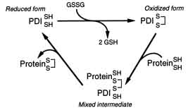
Cellular Component
The folding of secretory proteins occurs within the lumen of the endoplasmic reticulum. Consequently, the yeast protein PDI1, is also found in this region (Norgaard 2001).
Related Genetic Disorders
Within the Saccharomyces cerevisiae genome, there are at least four other genes with homology to PDI1 (figure 4) that produce PDI-like proteins.
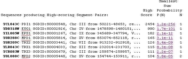
Figure 4 PDI-like Proteins. This figure illustrates the results of a BLASTp search, using the PDI1 protein as the query sequence. The underlined results represent proteins with PDI-like protein properties. Image from: Gish 2004. Permision pending.
More Protein Information from the Web
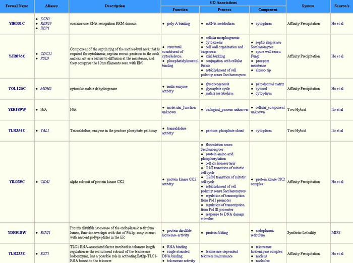
Figure 5 Proteins with which PCI1 is Known to Associate. This table illustrates the variety of proteins, representing a spectrum of processes, with which PDI1 associates. This variety leads one to assume that PDI1 may have broad-reaching effects, all of which are not yet fully understood. Image from: http://biodata.mshri.on.ca/. Permission pending.
A conserved domain search for the PDI1 sequence resulted in hits for thioredoxin, thiol-disulfide isomerase, and thiol:disulfide interchange domains (figure 6).
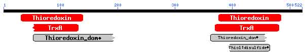
Figure 6 Conserved Domain Search for PDI1 Protein. This image shows conservation in the thioredoxin-like regions. Image from: http://www.ncbi.nlm.nih.gov/. Permission pending.
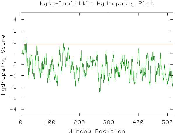
The Kyte-Doolittle hydropathy plot results (shown below) indicate the possibility of two transmembrane regions within the protein.
The PREDATOR in silico prediction for the secondary structure of PDI1 is shown below (figure 8).
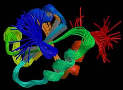
While there was no match for PDI1 within the protein database, a PDB-Homology search yielded 1mek, a human protein disulfide isomerase with 42% identity match and a 33% positive score < http://db.yeastgenome.org>. This protein is shown below.
My Favorite Non-Annotated Yeast ORF: YCL047C
Chromosomal Location
The YCL047C ORF is located within a small region of Saccharomyces cerevisiae chromosome 3, as shown below.
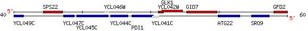
Figure 10 Chromosomal Location of YCL047C . This image shows the location of YCL047C on the Crick strand of yeast chromosome 3 (coordinates 44437- 43661). Also visible is the annotated PDI1 gene (right). Image from: http://db.yeastgenome.org/ (Cherry 1997). Permission pending.
Protein Information from the Web
The YCLO47C ORF is 777 bp long. To view the complete sequence, click http://db.yeastgenome.org/.
The YCL047C hypothetical protein is composed of 258 amino acids and has a molecular weight of 29,673 Daltons. To view the complete amino acid sequence, click http://db.yeastgenome.org/.
BLASTp results for the hypothetical protein sequence yield a number of results (the best hits are shown below).
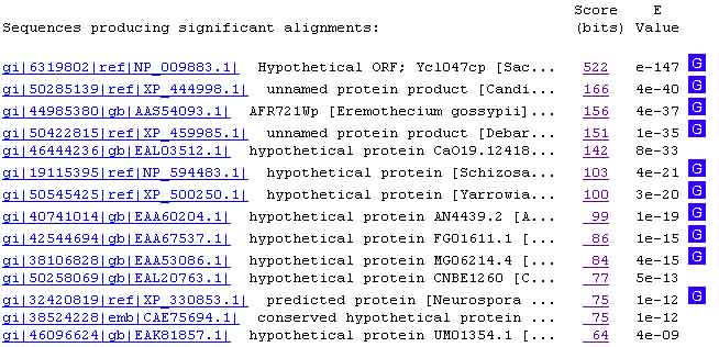
Figure 11 YCL047C-like Proteins. This figure illustrates the results of a BLASTp search, using the YCL047C hypothetical protein as the query sequence. Image from: http://www.ncbi.nlm.nih.gov/. Permission Pending.
While most of the strongest hits correspond to hypothetical proteins (in a variety of animals), the third hit is for the AFR721Wp protein in Eremothecium gossypii. Within this protein, there is a cytidylyltransferase conserved domain. Cytidylyltransferases form the critical intermediates in the biosynthesis of lipids and complex carbohydrates (Weber 1999).
A conserved domain search of the YCL047C sequence resulted in only one hit – the nicotinic acid mononucleotide adenylyltransferase domain, used in coenzyme metabolism. These results show a 98% alignment between the NadD region and that of YCL047C.
![]()
Figure 12 Conserved Domain Search for YCL047C hypothetical ORF. This image shows conservation of the nicotinic acid mononucleotide adenylyltransferase domain. Image from: http://www.ncbi.nlm.nih.gov/. Permission pending.
It is also interesting to note that YCL047C’s closest homolog (excluding hemiascomycetes) is a hypothetical protein within the Schizosaccharomyces pombe genome, called SPAC694.03 (figure 13). However, as this is also a hypothetical protein, the study of this homolog provides few insights. Yet, it is important to note that SPAC694.03 also contains a cytidylyltransferase conserved domain.
![]()
Figure 13 SPAC694.03, YCL047C's Closest Homolog. This figure illustrates that the SPAC694.03 hypothetical protein shares 31.5% identity with our YCL047C hypothetical protein. This makes SPAC694.03, found in Schizosaccharomyces pombe, the closest homolog of the YCL047C hypothetical protein. Image from: http://mips.gsf.de/. Permission Pending.
The Kyte-Doolittle hydropathy plot results (shown below) indicate the possibility of two transmembrane regions within YCL047C.
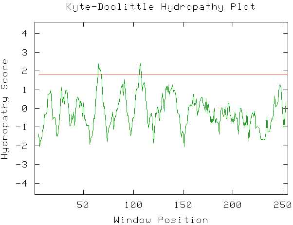
Figure 14 Kyte-Doolittle Hydropathy Plot of YCL047C. This plot predicts the presence of one transmembrane domain near amino acid 65 and a second transmembrane domain near amino acid 108. Image from: http://occawlonline.pearsoned.com/bookbind/pubbooks/bc_mcampbell_genomics_1/medialib/activities/kd/kyte-doolittle.htm. Permission granted by AM Campbell, PhD.
The PREDATOR in silico prediction for the secondary structure of the YCL047C hypothetical protein is shown below.
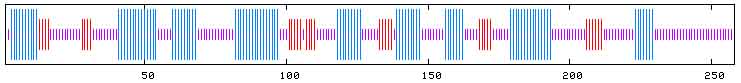
Figure 15 PREDATOR prediction for YCL047. Red areas indicate extended strand regions. Blue areas indicate alpha helices. Purple regions indicate areas of random coil. According to the PREDATOR prediction, 50% of this molecule would show a random coil patter, 37.21% would form alpha helices, and the remaining 12.79% would manifest in regions of extended strand. Image from: http://npsa-pbil.ibcp.fr/. Permission pending.
Disorders
References
Cherry JM, et al. Genetic and physical maps of Saccharomyces cerevisiae. Nature 1997 May 29; 387: 67-73.
Dolinski K, et al. 2004. Saccharomyces Genome Database. <http://www.yeastgenome.org>. Accessed 2004 Oct 4.
Giaever G, et al. Functional profiling of the Saccharomyces cerevisiae genome. Nature 2002 Jul 25; 418 (6896): 387-91.
Gish W. 2004. WU-Blast. <http://blast.wustl.edu>. Accessed 2004 Oct 4.
Givol D, et al. Disulfide interchange and the three-dimensional structure of proteins. Proceedings of the National Academy of Science USA 1995; 53: 676-684.
Heyer L, Johnson S, McCord RP, and Robinson L. Kyte Doolittle Hydropathy Plot. <http://occawlonline.pearsoned.com/bookbind/pubbooks/bc_mcampbell_genomics_1/medialib/activities/kd/kyte-doolittle.htm>. Accessed 2004 Oct 4.
Holst B, et al. Active Site Mutations in Yeast Protein Disulfide Isomerase Cause Dithiothreitol Sensitivity and a Reduced Rate of Protein Folding in the Endoplasmic Reticulum. Journal of Cell Biology 1997 Sep 22; 138(6): 1229-1238.
[IBCP] Institut de Biologie et Chimie des Proteines. 2001 Jan 23. Pole BioInformatique Lyonnais: Network Protein Sequence Analysis. <http://npsa-pbil.ibcp.fr/cgi-bin/npsa_automat.pl?page=/NPSA/npsa_preda.html>. Accessed 2004 Oct 7.
[MIPS] Munich Information Center for Protein Sequences. 2003 Nov 3. CYGD: Comprehensive Yeast Genome Database. <http://mips.gsf.de/genre/proj/yeast/index.jsp>. Accessed 2004 Oct 7.
Mount Sinai Hospital. 2003. Yeast Grid. <http://biodata.mshri.on.ca:80/yeast_grid/servlet/SearchPage>. Accessed 2004 Oct 7.
[NCBI] National Center for Biotechnology Information. 2004 Sep 8. NCBI Homepage. <http://www.ncbi.nih.gov>. Accessed 2004 Oct 7.
Norgaard P, et al. Functional differences in yeast protein disulfide isomerases. The Journal of Cell Biology 2001 Feb 5; 152(3): 533-562.
[RCSB] Research Collaboratory for Structural BioInformatics. 2004 Oct 5. RCSB PDB: Protein Data Bank. <http://www.rcsb.org/pdb>. Accessed 2004 Oct 7.
Weber CH, et al. A prototypical cytidylyltransferase: CTP:glycerol-3-phosphate cytidylyltransferase from Bacillus subtilis. Structure 1999; 7(9):1113-1124.
