This is a series of three animated GIF files that helps you
understand photosynthesis. This work was taken from the Virtual
Cell. Be patient and allow these three animation files enough
time to fully load onto your computer. Once loaded, each will
cycle slowly and repeatedly.
(For more information on the Virtual Cell, go to that site or
click here for details and credits.
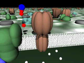
Figure 1. This shows light hitting photosystem II (blue-green in color) andphotosystem I (dark green color). Also shown are cytochrom b6/f (gray color) , and ATP-synthase (brown color).

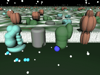
Figure 2. This animation shows PSII absorbing light and consuming water (1 blue-green oxygen and 2 white hydrogens) to replace the electron holes produced by the photooxydation of chlorophyll a. The electrons are incorporated into chlorophyll and oxygen (blue-green) is produced as a waste product and the hydrogen ions are retained to increase the concentration of hydrogen ions inside the thylakoid. Later in the animation, an electron carrier (yellow) is shown to move on to cytochrome b6/f and carry with it two more hydrogen ions to be added to the growing number of hydrogens in the thylakoid space.
Figure 3. This animation shows the consumption of ADP (1 blue plus 2 red phosphates) and a phosphate (1 red) to produce ATP (1 bue and 3 reds). This synthesis is driven by the proton-motive force which is depicted as white spheres leaving through the ATP-synthase.
The Virtual Cell. is an excellent place to explore and we strongly encourage you to click around. Due to the design of the virtual cell, it is difficult to give directions to these particular animations so we have collected them in one page for simplicity.
Project description and credits
Virtual Cell is being developed by Matej Lexa using POVRAY to generate 3-D images and PERL to write the script for navigation, with support from University of Illinois, namely SCALE and Prof. Richard Crang. The author has also produced a multimedia textbook for teaching photosynthesis and a searchable database of first names. He also has his own homepage. Some of the texts used here were adapted from:
A. B. Novikoff, E. Holtzman (1976) Cells and Organelles, 2nd ed. Holt, Rinehart and Winston, New York, 400 pp.
T. L. Lentz (1971) Cell Fine Structure. An Atlas of Drawings of Whole-Cell Structure. W. B. Saunders Company, Philadelphia, 437 pp.
Paul Thiessen from University of Illinois was kind to provide me with a POV-Ray DNA model made with his PovChem program. He has a nice page with related stuff on the Web.
Last major update: Jan 30, 1997.
![]()
![]()
©
Copyright 2000 Department of Biology, Davidson College, Davidson,
NC 28036
Send comments, questions, and suggestions to: macampbell@davidson.edu