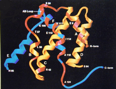
Figure 1: Domain structure of IFN-g
Residues 1-84 are shown in yellow, and 85-132 in blue. The residues
that contribute to IFN-g:receptor binding interface are highlighted in
red.
(Image produced by Walter et al. 1995, published in Nature)
Location on this page is pending approval by the authors