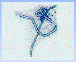
Image of the Ebola virus, courtesy of WHO
source: http://www.who.int/emc/diseases/ebola/ebolapic.html
see Copyright Information
* This web page was produced as an assignment for an undergraduate course at Davidson College *

Image of the Ebola virus, courtesy of WHO
source:
http://www.who.int/emc/diseases/ebola/ebolapic.html
see Copyright
Information
The Ebola (EBO) virus is a member of the family filoviridae which is composed of non-segmented negative strand RNA viruses. There are four known subtypes of Ebola: Zaire, Sudan, Reston, and Ivory Coast (EBO-Z, EBO-S, EBO-R, and EBO-IC, respectively). EBO-Z is the most virulent subtype, with a 77% mortality rate in its most recent positively identified outbreak in Zaire in 1995 (Sanchez et al., 1996). However, not all subtypes are as virulent. EBO-IC is pathogenic in primates, but only caused a single nonfatal human infection. Likewise, EBO-R is only pathogenic in primates. However, four exposed humans became seropositive, but failed to develop any symptoms of infection (Formenty et al., 1999).
Virulent strains of Ebola cause a condition known as hemorrhagic fever (HF). Ebola HF is a particularly severe form of HF, the symptoms of which are: high fever, severe hemorrhaging, and shock (Mühlberger et al., 1999). The exact means through which HF is achieved are highly diverse, quite impressive, and poorly understood. Comparison studies of the humoral response of fatalities to survivors have provided an insight into the havoc an Ebola infection creates. Ebola-specific IgG and IgM were found early in the infection in all survivors. However, only in one third of fatalities was IgM detected and never was any IgG detected. The survivors all possessed IgG against viral nucleoprotein. Some also produced IgG against other viral proteins, but no IgG was specific for the transmembrane glycoprotein (GP). T cell functions were examined through studies of the cytokines and proteins released during the course of the infection. Differences in cytokine and protein upregulation between survivors and fatalities were observed. Fatal cases of Ebola HF are characterized by upregulation of gamma interferon (IFN-g), Fas, Fas ligand (FasL), and perforin and no upregulation of CD28 mRNA synthesis, a receptor for the co-stimulatory signals to activate T cells. As the symptoms progressed massive amounts of IFN-g and secreted (s)FasL were produced, indicating the activation of many cytotoxic (CD8+) T cells and other cytolytic cells. However, in survivors during the symptomatic phase, the levels of IFN-g, Fas, FasL, perforin, and CD28 mRNA were virtually the same as endemic controls. Upregulation of these molecules indicated a period of antigen clearance and clinical recovery. As in the fatalities, this upregulation is indicative of cytotoxic T cell activation (Baize et al., 1999).
The massive cytolytic cell activation in fatalities was followed by the complete loss of T cell related CD3 and CD8 mRNA. IFN-g levels also correspondingly decreased. Bcl-2, an apoptosis inhibitor, and 41/7 nuclear matrix protein (NMP), a protein released during apoptosis, decreased and increased synthesis, respectively. 41/7 NMP levels increased for the last five days prior to death, which is indicative of massive apoptosis of cells from diverse origins, but especially T cells. The evidence suggests that the apoptosis observed in fatalities results from excessive activation of T cells which may lead to downregulation of the T Cell receptor (Baize et al., 1999). Furthermore, fatalities have also been observed to upregulate IL-2, a T cell proliferation factor, as well as IFN-g. Interestingly, IL-6 upregulation was not observed during the infection. Endothelial cells are a source of IL-6, suggesting that these cells do not respond to cytokines during Ebola infection (Villinger et al., 1999).
The responses of endothelial cells have been shown to be defective
during an Ebola infection. Seventy-two hours post infection
(p.i.) the MHC class I levels on endothelial cells are 50% of
normal. IFN-g causes a three fold increase in MHC I expression
in normal endothelial cells; however, during an Ebola infection
IFN-g has no effect on the decreasing MHC I expression.
The effects of IFN-a are also blocked during the infection.
Conversely, IL-1b can induce IL-6 production in endothelial cells.
Clearly, Ebola (in this case EBO-Z) does not block all signal
transduction or de novo protein synthesis. A potential point
of inhibition occurs at the janus kinase (Jak)-1 or signal transducers
and activators of transcription (STAT)-1a pathway (Harcourt et
al., 1999).
Ebolaís effect on endothelial cells is surmised from
the ability of Marburg, a related filovirus, to directly infect
endothelial cells. Marburg has been clearly shown to replicate
in endothelial cells during the infection. At the terminal
phase of Marburg HF, shock is caused as a result of direct lysis
of endothelial cells by the Marburg virus, massive release of
inflammatory mediators by unspecific (phagocytosis) or specific
(viral replication) activation of inflammatory cells, and oxidant
injury or unspecific immune responses (Schnittler et al., 1993).
The severe shock associated with Ebola HF can be attributed in
part to the destruction of the barrier created by the endothelial
cells. In Ebola infections TNFa is thought to be a primary
cause of the shock associated with Ebola HF. TNFa induces
several changes in endothelial cells. TNFa causes increases
in endothelial permeability, loss of anticoagulant activity, and
expression of adhesion molecules that recruit platelets and leukocytes.
The apparent upregulation of TNFa is caused by macrophages and
monocytes that are activated to release TNFa upon infection by
Ebola (Feldmann et al., 1996).
Macrophages and monocytes are fully susceptible to infection
and replication of filoviruses. Moreover, macrophages and
monocytes serve as a vehicle for dispersion of viral particles.
The viral particles undergo intracellular budding and accumulate
in vacuoles, thus hidden virus is spread and evades detection
by the immune system (Feldmann et al., 1996). In one study
macrophages were the only cells that contained Ebola viral antigen
in its cytoplasm. The cytoplasmic viral particles were especially
concentrated in the spleen, liver, and circulating cells (Wyers
et al., 1999). It is thought that the combination of endothelial
infection due to viral replication and cytokines released from
macrophages and monocytes provides the conditions conducive to
a distinct proinflammatory endothelial phenotype. The result
of which is a coagulation cascade that leads to disseminating
intravascular coagulation (DIC), a complicating condition that
arises in the final stages of the disease. The final stages
of shock and hemorrhagic manifestations potentially result from
uncontrolled virus-induced lysis of macrophages and monocytes.
The lysed macrophages and monocytes release massive amounts of
secretory products, which may reduce the integrity of the microvascular
wall (Feldmann et al., 1996).
In order for Ebola to cause infection, it must be capable
of entering cells. Ebola fusion peptides have been shown
to destabilize lipid vesicles which allows membrane fusion.
The fusion protein is a glycoprotein (GP) that is not inert, but
causes perturbations of the bilayer that results in membrane fusion.
The action is thought to be analogous to the HIV fusion peptide.
Fusion was found to only occur in the presence of calcium cations
and phosphatidylinositol in the bilayer (Ruiz-Argüello et
al., 1998). The fusion protein, GP, forms homotrimers in
the viral membrane. Each GP subunit is heterodimer comprised
of GP1 and GP2 (Sanchez et al., 1998). The gene that codes
for GP is differentially transcribed by the RNA dependent RNA
polymerase (L). As a result two different glycoproteins
are produced, sGp, a truncated secreted form, and GP, the membrane
form. GP is then split by furin or subtilisin-like eukaryotic
proteases all of which are found ubiquitously, but especially
in hepatocytes and endothelial cells. Furin is known to
cleave at the site RXRR in a peptide. The Ebola subtypes
EBO-Z, S, and IC express this sequence and are cleaved.
However, EBO-R expresses KQKR. As a result furin cleavage
is less favorable. It seems possible then that subtype virulence
could be determined by the amino acids sequence at the cleavage
site (Volchkov et al., 1998). GP is cleaved into GP1 and
GP2, which forms a heterodimer through disulfide bonding.
GP1 is able to dissociate from the heterodimer and be released.
sGP and GP1 are phagocytosed by macrophages and other APCís
when in secreted form. Those peptides are then presented
on MHC class II, which elicits a lytic response by CD4 T cells,
a result also observed with GPís of HIV, VSV and influenza
virus. Endothelial cells may also be subject to lysis by
CD4 T cells when expressing sGP or GP1 in MHC II in addition to
destruction by viral replication. These secreted GPís
may also bind antibodies, inhibiting a humoral response (Volchkov,
1999). Interestingly, lymphoid lineage cells are unable
to be infected by Ebola. The GP fusion is possibly inhibited
by lymphoid cells, or cells of that lineage lack the necessary
recpetor (Wool-Lewis and Bates, 1998).
GP2 serves not only to facilitate fusion of membranes, but
also may have an immunosuppressive domain. The terminal
region of the GP2 has high homology with p15E-related GP of oncogenic
retroviruses. This motif inhibits mitogen mediated blastogenesis
of lymphocytes, decreases monocyte chemotaxis and macrophage infiltration,
and decreases proliferation of mononuclear cells. However,
the true function of this motif in Ebola is unknown (Volchkov,
1999).
The sGP also has a suspected function when in its secreted
homodimeric form. sGP is known to inhibit early neutrophil
activity. The mechanism for this interaction is not clear,
but is related to the IgG receptor IIIb (FcgRIIIb, CD16) that
is expressed solely on neutrophils. SGp has been shown not
to directly bind to CD16, but crosslinking of CD16 may induce
the sGP receptor, thereby inactivating the neutrophil (Marayuma
et al., 1998).
Ebola not only attacks the immune system by horribly disrupting
its signaling pathways and inflammation response, but also destroys
the tissues largely responsible for immune function. Two
to three days p.i. microcirculatory disruptions are present, especially
in liver and spleen. Macrophages, stromal, and dendritic
cells swell in the lymphoid tissue. Mitosis and involution
of the lymphoid follicle are also blocked. Four to six days
p.i. the severest damage is observed the red pulp of the spleen.
The walls are completely destroyed and is accompanied by hemorrhaging.
The stroma and macrophages have become necrotic. Similarly,
in the white pulp and in the lymph follicles of the respiratory
and GI tracts, macrophages and stromal cells have become necrotic
and lymph tissue exhibits increased lymphoid depletion.
The pathomorphologic appearance of the lymphoid tissue is characterized
by swelling of the vascular epithelium, thrombosis, fibrin deposits,
and diapedesis of erythrocytes (Ryabchikova et al., 1999).
Clearly, Ebola has developed many tricks to defeat our immune system. Surviving Ebola HF until recently required a lot of luck and an ìÖ orderly and well-regulated humoral and cellular immune responses,î (Baize et al., 1999). An outbreak of EBO-Z in Kikwit, Democratic Republic of the Congo, brought about a small success in defeating infection. Serum from five convalescent patients was transfused in desperation into 8 patients exhibiting symptoms of Ebola HF. All eight were very ill, four of which had hemorrhagic manifestations, and 2 of which became comatose. The transfusions saved all but one life. This is a drop in mortality rate from 80% to 12.5%. However, other attempts with transfusion of convalescent serum have proven much less successful (Mupapa et al., 1999). Other attempts at passive immunity have been tried. The use of equine antibodies has met with limited success (Kudoyarova-Zubavichene et al., 1999). In a murine model, subjects were vaccinated against sGP or GP. Some did develop protective immunity, always in the form of an IgG2a antibody. Whereas, IgG 1 and IgG3 were never protective. This study demonstrates that it is possible to elicit protection by vaccination or produce antibodies for therapeutic use (Wilson et al., 2000). An antiviral therapy approach holds great promise. S-Adenosylhomocysteine hydrolase inhibitors block transmethylation ractions through a feedback mechanism. Without the transmethylation of the RNA, no mRNA replication can occur. If administered at the early symptomatic phase, a 90% success rate has been achieved. If administered later, the success rate drops considerably and toxicity problems arise (Huggins et al., 1999). The most promising treatment is genetic immunization. A gene for a particular viral peptide is inserted into the host genome, in this case sGP, GP, or NP. The inserted gene is expressed, allowing the body to mount an immune response. Immunization against sGP and GP provided the best immunity, 100% at two months and 60% at four months. NP had a survival rate of 100% after two months and 0% after four months. SGP and GP are also have few nucleotide polymorphisms, making them great epitopes for immunization (Xu et al., 1998).
Despite Ebolaís surge into the mainstream of virology,
little is really known about this pathogen. The animal that
serves as the viral reservoir is still unknown. Many species
can serve as intermediate hosts, especially non-human primates,
but Ebola is pathogenic to them as well. Some feel that
the reservoir may be a species of solitary bat or it may be arthropod-borne
(Monath, 1999). Bats experimentally infected with Ebola
do not die, supporting the bat reservoir theory. Another
theory is that filoviruses evolved from plant viruses (WHO, 1997).
Traces of non-pathogenic or defective Ebola viral particles have
been found in about 3% of the most common species in and around
areas of outbreaks, but none of these are capable of starting
an infection (Hagmann, 1999). Furthermore, in some human
populations the number of Ebola seropositive individuals exceeds
30%. Clearly, non-pathogenic variants occur relatively frequently
(Monath, 1999), but then outbreaks start usually from one infection,
why? Although we are a long way from discovering all of
Ebolaís secrets, our knowledge is already able to reap
potential benefits. Because of Ebola GPís ability
to bind to endothelial cells, researchers are able to target therapeutic
genes specifically at these cells. One such application
could deliver growth factors to stimulate the growth of new blood
vessels in place of damaged vessels, treating cardiovascular disease
(Wickelgren, 1998).
References
Baize S, Leroy EM, Georges-Courbot MC, Capron M, Lansoud-Soukate J, Debré P, Fisher-Hoch SP, McCormick JB, Georges AJ. Defective humoral responses and extensive intravascular apoptosis are associated with fatal outcome in Ebola virus-infected patients. Nature Medicine 5(4):423-426.
Feldmann H, Bugany H, Mahner F, Klenk HD, Drenckhahn D, Schnittler HJ. 1996. Filovirus-induced endothelial leakage triggered by infected Monocytes/Macrophages. Journal of Virology 70(4):2208-2214.
Formenty P, Hatz C, Le Guenno B, Stoll A, Rogenmoser P, WidmerHuman A. 1999. Infection Due to Ebola Virus, Subtype Côte díIvoire: Clinical and Biologic Presentation. The Journal of Infectious Diseases 179(Suppl 1):S48-53.
Hagmann M. 1999. On the track of Ebolaís hideout. Science 286:654-655.
Harcourt BH, Sanchez A, Offermenn MK. 1999. Ebola virus selectively inhibits responses to interferons, but not to Interleukin-1b, in endothelial cells. Journal of Virology 73(4):3491-3496.
Huggins J, Zhang ZX, Bray M. 1999. Antiviral drug therapy of filovirus infections: S-Adenosylhomocysteine hydrolase inhibitors inhibit Ebola virus in vitro and in a lethal mousee model. The Journal of Infectious Disease 179(suppl 1):S240-247.
Kudoyarova-Zubavichene NM, Sergeyev NN, Chepurnov AA, Netesov SV. 1999. Preparation and use of hyperimmune serum for prophylaxis and therapy of Ebola virus infection. The Journal of Infectious Diseases 179(suppl 1):S218-223.
Monath TP. 1999. Ecology of Marburg and Ebola viruses: Speculations and directions for future research. The Journal of Infectious Diseases 179(suppl 1):S127-138.
Maruyama T, Buchmeier MJ, Parren P, Burton DR. Ebola virus, neutrophils, and antibody specificity. Science 282:845a.
Mühlberger E, Weik M, Volchkov VE, Klenk HD, Becker S. 1999. Comparison of the transcription and replication strategies of Marburg Virus and Ebola Virus by using artificial replication systems. Journal of Virology 73(3):2333-2342.
Mupapa K, Massamba M, Kibadi K, Kuvula K, Bwaka A, Kipasa M, Colebunders R, Muyembe-Tamfum JJ. 1999. Treatment of Ebola Hemorrhagic Fever with Blood Transfusions from Convalescent Patients. The Journal of Infectious Diseases 179(suppl 1):S18-23.
Ruiz-Argüello MB, Go?i FM, Pereira FB, Nieva JL. Phosphatidylinositol-dependent membrane fusion induced by a putative fusogenic sequence of Ebola virus. Journal of Virology 72(3): 1775-1781.
Ryabchikova EI, Kolesnikova LV, Luchko SV. 1999. An analysis of features of pathogenesis in two animal models of Ebola virus infection. The Journal of Infectious Disease 179(suppl 1):S199-202.
Sanchez A, Trappier SG, Mahy BWJ, Peters CJ, Nichol ST. 1996. The virion glycoproteins of Ebola viruses are encoded in two reading frames and are expressed through transcriptional editing. Proceedings of the National Academy of Science 93:3602-3607.
Sanchez A, Yang ZY, Xu L, Nabel GJ, Crews T, Peters CJ. 1998. Biochemical analysis of the secreted and virion glycoproteins of Ebola virus. Journal of Virology 72(8):6442-6447.
Schnittler HJ, Mahner F, Drenckhahn D, Klenk HD, Feldmann H. 1993. Replication of Marburg virus in human endothelial cells: a possible mechanism for the development of viral hemorrhagic disease. Journal of Clinical Investigations 91:1301-1309.
Villinger F,Rollin PE, Brar SS, Chikkala NF, Winter J, Sundstrom JB, Zaki SR, Swanepoel R, Ansari AA, Peters C. 1999. Markedly elevated levels of Interferon (IFN)-g, IFN-a, Interleukin (IL)-2, IL-10, and Tumor Necrosis Factor-a associated with fatal Ebola virus infection. The Journal of Infectious Diseases 179(Suppl 1):S188-191.
Volchkov VE, Feldmann H, Volchkova VA, Klenk HD. 1998. Processing of the Ebola virus glycoprotein by the proprotein convertase furin. Proceedings of the National Academy of Science 95:5762-5767.
Volchkov VE. 1999. Processing of the Ebola virus glycoprotein. Current Topics in Microbiology and Immunology 235:35-47.
WHO. 1997 Sept. Ebola Hemorrhagic Fever <http://www.who.int/inf-fs/en/fact103.html> Accessed 2000 Apr 21.
Wickelgren I. 1998. A method in Ebolaís madness. Science 279:983-984.
Wilson JA, Hevey M, Bakken R, Guest S, Bray M, Schmaljohn AL, Hart MK. 2000. Epitopes involved in antibody-mediated protection from Ebola virus. Science 287:1664-1666.
Wool-Lewis RJ, Bates P. 1998. Characterization of Ebola virus entry by using pseudotyped viruses: identification of receptor-deficient cell lines. Journal of Virology 72(4):3155-3160.
Wyers M, Formenty P, Cherel Y, Guigand L, Fernandez B, Boesch C, Le Guenno B. 1999. Histopathological and Immunohistochemical Studies of Lesions Associated with Ebola Virus in a Naturally Infected Chimpanzee. The Journal of Infectious Diseases 179(suppl 1):s54-59.
Xu L, Sanchez A, Yang ZY, Zaki SR, Nabel EG, Nichol ST, Nabel
GJ. 1998. Immunization for Ebola virus infection.
Nature Medicine 4(1):37-42.
Related Sites
The Centers for Disease Control and Prevention
Virology Institute at Marburg, Germany
Return To Immunology Home
Page of
Charles Haines
Return
To Immunology Main Page
Send comments, questions, and suggestions to: chhaines@davidson.edu