Molecular Method Page
Timothy S. Deeb
Molecular Biology
University of California researchers B. Albert Griffin, Stephen R. Adams, and Roger Y. Tsien (1998) published results on their experimental method for "Specific Covalent Labeling of Recombinant Protein Molecules" within living cells. The researchers studied covalent bond formation between "organo-arsenicals and pairs of thiols." The binding of trivalent arsenic compounds to thiol proteins with pairs of cysteines or lipoic acid causes much of the toxicity of arsenic compounds, but can be "completely reversed by small vicinal dithiols" as antidotes. The researchers used this characteristic of the antidotes to begin their efforts to design a peptide domain and a ligand that would form even tighter bonds. In the presence of excess antidote, this ligand could bind the selected peptide domain without toxicity to other proteins. This method utilizes the tight-binding between a small peptide "receptor domain composed of as few as six natural amino acids that could be genetically incorporated into proteins of interest, and a small (<700-Dalton), synthetic, membrane-permeant ligand" that is nonfluorescent "until it binds with high affinity and specificity" to the target domain.
From fourteen biarsenical ligands, the researchers were able to create one successful ligand. The researchers prepared "[4',5'-bis (1,3,2-dithioarsolan-2-yl) fluorescein," or "FLASH-EDT2, [fluorescein arsenical helix binder, bis-EDT adduct] by the process of transmetallation of fluorescein mercuric acetate, adding EDT (1,2-ethanedithiol) to purify. The initially nonfluorescent product developed bright fluorescence with the addition of a tetracysteine peptide to displace the EDT. The researchers suggest
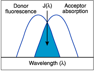
the small size of EDT probably permits rotation of the aryl-arsenic bond and excited state quenching by vibrational deactivation or photoinduced electron transfer, whereas the peptide complex may evade such quenching because its more rigid conformation should hinder conjugation of the arsenic lone pair electrons with the fluorescein orbitals (Griffin, Adams, and Tsien 1998).Griffin, Adams, and Tsien (1998) went on "to test the membrane permeability of FLASH-EDT2 and the specificity of the FLASH-peptide interaction in live mammalian cells" by genetically fusing this peptide (after changing the W to an A) to the "COOH-terminus of an enhanced cyan fluorescent protein (ECFP)." This fusion protein was then transiently expressed in HeLa cells (a mammalian line of cells). The researches demonstrated the following: 1) In the absence of FLASH-EDT2, "some cells were brightly fluorescent at the ECFP emission maximum (480 nm) and only very dimly fluorescent at 635 nm, at which ECFP barely emits." 2) Addition of FLASH-EDT2 and EDT to cells "expressing ECFP increased their fluorescence at 635 nm more than threefold, whereas the fluorescence of ECFP at 480 nm declined by >70%". The researchers used "fluorescence resonance energy transfer (FRET)" to demonstrate the formation of the ECFP-Cys4 peptide-FLASH complex.
Limitations of the Labeling Method
The researchers mentioned one limitation for the binding of FLASH to the tetracysteine domain (CCXXCC): the "thiols must be able to reach an alpha-helical or other conformation able to form two pairs that grip the arsenics like pincers, yet the thiols must not be disulfide-bonded or tightly chelated to a metal" (Griffin, Adams, and Tsien 1998).
Advantages of the Labeling Method
Being able to control the onset and reverse the binding (and therefore the toxicity) and labeling makes this a process of special significance. When the EDT antidote was administered, the HeLa cells remained viable for up to four hours; without the EDT, the labeling substance was highly toxic. Using EDT with the arsenic permits the use of the process in live cells. (Griffin, Adams, and Tsien 1998).
FLASH acts as an acceptor; therefore, it allows for a comparison of donor emission before and after labeling with FLASH, which is one of the keys to its specificity.
Although Griffin, Adams, and Tsien point out that while not all native proteins which contain the core motif CCXXCC will be likely to bind FLASH, "endogenous competing proteins or ligands are clearly rare enough in mammalian cells to permit easy detection of transfected proteins over background" (1998). According to the researchers, derivatives of FLASH, whether luminescent, magnetic, fluorescent, or photochemically reactive, should be able to label proteins with the CCXXCC motif.
This molecular method
combines high affinity and specificity, easy reversibility, easy modification of the ligand, small size of the peptide domain, physiological compatibility, and fluorescence indication that binding has occurred (Griffin, Adams, and Tsien 1998).The advantages of this molecular labeling method over other methods such as GFP and immunofluorescence labeling include the following: 1) this method utilizes small molecular components which are more permeable and contribute less mass, 2) this labeling method has an increased selection of attachment sites on the target protein, 3) this labeling method uses a ligand which is nonfluorescent until it binds its target peptide domain, 4) this method permits targeting of recombinant proteins inside live cells.
Reference
Griffin BA, Adams SR, and Tsien RY. Specific covalent labeling of recombinant protein molecules inside live cells. Science 1998:281:269-272.
Figure 1. This is a schematic representation of the FRET spectral overlap integral.