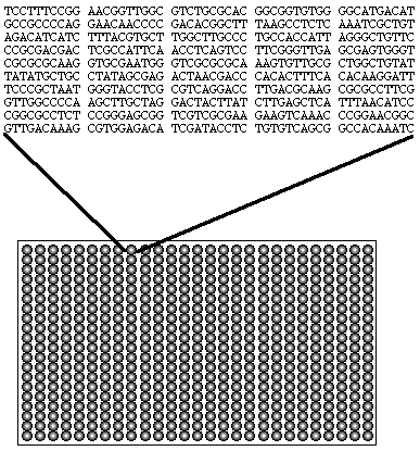
Fig.1.2 This figure is a cartoon illustrating an array of DNA snippets on a chip. The top portion depicts a possible nucleotide sequence for the DNA segment immobilized in the position indicated.
Thus far, the Molecular Biology
(Bio. 304) class at Davidson College has studied many different methods
including Polymerase Chain Reaction (PCR), hybridization, gel electrophoresis,
and Southern and Northern blots. Each of these methods is or can
be used in the various steps of the DNA Microarray method. However,
no method we have yet studied even comes close to the efficiency, amount
of data, and endless possibilities of DNA Microarrays. There are
actually many different microarray methods that have been developed and
more that are in the process of being developed. However, each array
system seems to follow the same basic method. Therefore, the general
method is described below with the assistance of two different examples.
Further down on the page you will find a discussion of the applications
of DNA Microarrays followed by a list of related links. Enjoy!
General Method:
First, various "snippets"1 of DNA of the organism or
cells being investigated, are arranged in a certain array and immobilized
on chips, or slices, of glass or silicon which tend to be about 2 or 3
centimeters in width.1
Fig.1.2 This figure is a cartoon illustrating an array of DNA snippets on a chip. The top portion depicts a possible nucleotide sequence for the DNA segment immobilized in the position indicated.
Next, two different samples of DNA are prepared isolation fragmentation using methods such as PCR and gel electrophoresis.3,4 In the case of Elizabeth A. Winzeler et. al, who were investigating the allelic variation of the yeast genome, these samples were genomic DNA from two different strains of yeast.3 In experiments where gene expression is being investigated, generally the cells being studied are exposed to two different conditions. The mRNA is then harvested from the cells and cDNA is then produced. This cDNA is then prepared as mentioned above. (Don't forget, there are two different samples of cDNA!) Each of the two DNA samples is then labeled with a different fluorescent tag.1-4
The above DNA samples are then introduced to the prepared chip at which point hybridization, base pairing, occurs between the DNA immobilized on the chip and the fluorescently tagged DNA. The chip is then washed and stained if necessary, depending on the tag being used.1-4
At this point, the chip is scanned to detect the fluorescence at the various positions on the chip.1-4
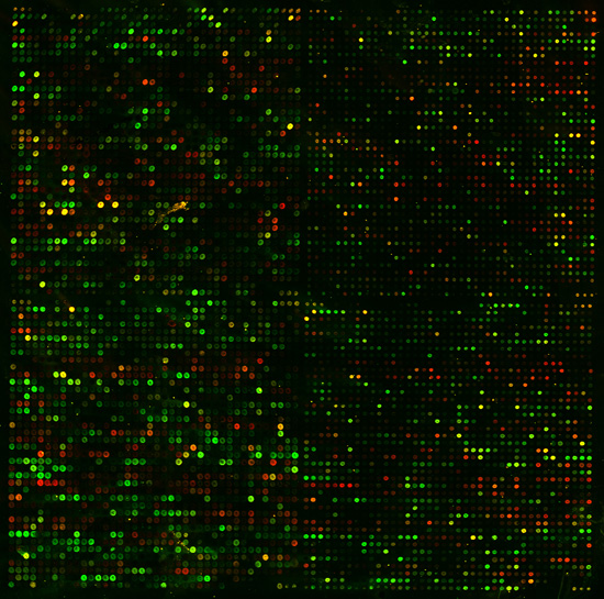
Fig.2.4 This figure, courtesy of Pat Brown's lab, is the scanned image of a DNA Microarray chip containing the entire yeast genome. Spots in which only green or red appear are positions in which only one of the probes (tagged DNA described above) base paired to the chip. In general, spots that are yellow are positions at which both probes base paired to the chip to some degree.
Once the chip has been scanned, it must be analyzed. The exact analysis that is done in part depends on the particular experiment. However, in general The ratios of the two different fluorescence present for each spot is measured. Then comparisons may be made between different chips. For example, in the case of the experiment in which allelic variation of the yeast genome was being investigated, two different arrays were used and then compared which "revealed hybridization differences for the two strains."3 In cases in which gene expression is being investigated, the colors may next be linked to expression followed by the grouping of "genes with similar profiles."2
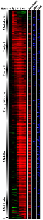
Fig.3.2 This figure is an example of Microarray results that have been analyzed such that the colors were linked with expression and then similar gene profiles were grouped together. The data was then organized into the figure above. More specifically, this particular figure represents the data collected from an experiment on the budding of yeast. (For more information please visit Dr. Campbell's Microarray site.)
Applications:
According to the director of the National Human Genome
Research Institute (NHGRI), Francis Collins, the application of DNA Microarrays
"is just exploding in all kinds of directions."1
This versatile method has already been used by researchers to compare the
effects of different drugs on gene expression1
as well as to scan the yeast genome for allelic variations.3
In addition, Collins and other researchers at the NHGRI have utilized Affymetrix
chips "to detect mutations in the familial breast cancer gene BRCA I
in
subjects at risk for the disease."1
Using a different set of arrays, Jeffrey Trent and others investigated
the effects of radiation treatment on "gene expression in cancer cells."1
What's more, researchers like Pat Brown of Stanford, one of the leaders
in microarrays, are coming up with more data than they know what to do
with! In addition to these current applications, researchers are
harboring high hopes for the possibilities of microarrays in future work.
For example, some researchers hope that DNA arrays will illustrate
"unique gene-expression patterns" that will mark the onset of diseases
ranging from cancer to osteoporosis to Alzheimer's' to heart disease.1
Another possibility for arrays that researchers are hoping for is use in
measuring the effectiveness of drugs used to fight HIV. DNA Microarrays
may also allow for patient-specific medications based on the patients unique
genetic makeup as well as being a method for sequencing genes. The
implications of the above possibilities for diagnostic companies, pharmesuticals,
and the medical community in general are huge. "[Researchers] expect that
understanding the genes active in disease will spawn a new generation of
therapeutic drugs that treat underlying causes rather than symptoms."1
Further, Wei Zhang of M.D. Anderson Cancer Center states, "With the new
technology, we can analyze a huge number of genes at the same time.
That provides for a new era of diagnostics and therapeutics."1
The experiments and possibilities mentioned above barely even skim the
surface of what has been done and can be done using DNA microarrays.
In the words of Collins, "The limits will not be found anytime soon."1
For more information on DNA Microarrays or the experiments mentioned above, please click on the links below:
"The Brown Lab" (Pat Brown, Stanford University, Ca.)
Dr.
Campbell's Microarray site (A. Malcolm Campbell, Davidson College,
NC, and Mary Lee
Ledbetter, College of the Holy Cross,
MA)
A
Sample of Published Microarray Experiments (A. Malcolm Campbell,
Davidson College,
NC, and Mary Lee Ledbetter, College
of the Holy Cross, MA)
For these and other valuable links:
"Home
Page for the Genome Consortium for Active Teaching (GCAT)" (A. Malcolm
Campbell,
Davidson College, NC, and Mary Lee
Ledbetter, College of the Holy Cross, MA)
2 Campbell, A. Malcolm and Mary Lee Ledbetter. 1999 Dec. 6. Methodology for Functional Genomics. <http://bio.davidson.edu/Biology/GCAT/ASCBpresentation/ASCB3.html> Accessed 2000 Feb. 20.
3 Winzeler, Elizabeth A. et al. 1998. Direct Allelic Variation Scanning of the Yeast Genome. Science 281: 1194-1197.
4 Brown, Pat. 2000
Feb. 8. The Brown Lab. <http://cmgm.stanford.edu/pbrown>.
Return to Davidson College Biology Department Home Page
Return To Biology Course Materials
![]()