The Genisphere 3DNA Method
This Genisphere, Inc. procedure is summarized here for academic purposes.
All questions, comments and ordering requests should be sent to the
people who make 3DNA: ![]()
The Genisphere 3DNA Method
This Genisphere, Inc. procedure is summarized here for academic purposes.
All questions, comments and ordering requests should be sent to the
people who make 3DNA: ![]()
3DNA is a nucleic acid detection
technique recently developed by Genisphere,
Inc. 3DNA is an efficient hybridization probe that allows for
easy detection of a very small amount of target nucleotide. You will
soon see why 3DNA is a highly effective technique for nucleic acid blotting.
Structure of the 3DNA Probe
The 3DNA technique can attribute its effectiveness to the structure of the 3DNA probe. While traditional probes for Northern and Southern blots consist of a single nucleotide sequence for target molecule detection,1 the 3DNA probe consists of several hundred target-specific nucleic acid sequences within a single molecule that can bind to the target nucleotide strand. This probe is called a dendrimer.
Assembly of the 3DNA Dendrimer
The 3DNA dendrimer consists
of several layers of the monomer shown below2 (Figure 1).
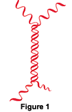
(Source for diagram is Genisphere)
This monomer consists of two ssDNA strands with a central region of complementary nucleotide sequence. The central region where the DNA is base-paired is referred to as the "waist" of the monomer, and extending from the waist are four unbonded "arms" of noncomplementary nucleic acid sequence.2
The 3DNA dendrimer is formed by joining several layers of these monomers. The arms of the monomer are designed to base-pair with the arms of other monomers in a precise fashion to produce several layers that interact to form a complete dendrimer.2 At the center of the dendrimer is a single "initiator monomer." The arms of the initiator unit base-pair with the complementary nucleotide sequence of the arms of four other monomers to form the first layer of the dendrimer (Figure 2A). The free arms extending from the first layer of the dendrimer then base-pair with complementary arms of a second layer of monomers (Figure 2B). A third layer is added, and then a fourth and final layer of monomers is added to complete the dendrimer (Figure 2C). In order to avoid degradation of the dendrimer, the monomer arms are ligated between each layer at each stage.2
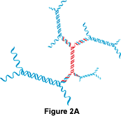
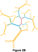
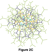
(Source for diagrams is Genisphere)
Using Oligonucleotides to Create a Functional 3DNA Probe With Labels
The complete dendrimer shown in Figure 2C has 324 exposed (unpaired) ssDNA arms extending from its outer layer. There are two types of arms, each with one of two nucleotide sequences: "a+" or "c+." Half of the arms are composed of the a+ sequence and the other half are composed of the c+ sequence. Therefore, there are 162 arms of each type extending from the dendrimer. The c+ arm is ligated to a labeled oligonucleotide (Figure 3A), while the a+ arm is ligated to a target-specific oligonucleotide probe for hybridization to the target nucleic acid sequence2 (Figure 3B).
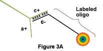
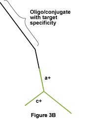
(Source for diagrams is Genisphere)
The completed 3DNA dendrimer after attachment of oligonucleotide probes and labeled oligonucleotides looks like this (Figure 3C):

(Source for diagram is Genisphere)
Advantages of 3DNA
Enhanced Sensitivity
Since the 3DNA dendrimer has 162 arms for the attachment of target-specific oligonucleotide probes, it has multiple binding sites for hybridization with a single target nucleic acid sequence. As opposed to a traditional probe that consists of only a single nucleotide sequence,1 the large dendrimer has 162 target-specific probes and thus has a greater likelihood of hybridizing to the target sequence. Therefore, 3DNA can hybridize successfully when only a small amount of target sequence is present. Moreover, because the 3DNA dendrimer is large in size and has multiple hybridization sites, addition of even a small amount of 3DNA can successfully detect the target sequence. Thus, very little 3DNA is needed to achieve a successful hybridization.2
In addition, 3DNA offers excellent signal strength from the oligonucleotide labeling. When an oligonucleotide probe on the 3DNA dendrimer hybridizes to a single target nucleic acid, its location is revealed by the 162 oligonucleotide labels also attached to the dendrimer. Therefore, the signal strength is enhanced 162-fold over a traditional probe with a single label. This provides a very intense signal that allows for detection of a very small amount of target nucleic acid.2
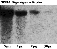
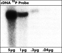
The multiple hybridization sites and the multiple labels of the 3DNA dendrimer result in approximately a 100-fold increase in sensitivity over traditional probes.3 The following diagram illustrates a hybridization of a portion of HIV genome in different quantities using a traditional oligonucleotide probe compared with a hybridization using 3DNA dendrimer.4 The strength of the signal from the oligonucleotide probe hybridized with a 40 pg DNA sample is roughly equivalent to the strength of the signal from the 3DNA dendrimer probe hybridized with a 156 fg DNA sample (Approximately a 256-fold increase in sensitivity).
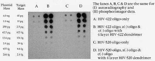
( Source for diagram is Polyprobe.
Genisphere 3DNA products were used in the hybridization.)
Versatility in Labeling Options
Several oligonucleotide labels are available from Genisphere, ranging from fluorescence to radioactive labeling.5
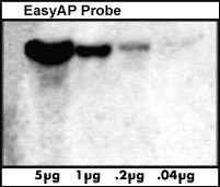
Detection of the follicle stimulating hormone receptor
gene from blots of the listed quantities of human genomic DNA
using 3DNA with the indicated label. (Source for
diagram is Genisphere)
Common Applications of 3DNA
Northern and Southern Blotting
The target detection abilities of 3DNA allow detection of extremely low quantities of DNA or mRNA target. 3DNA can detect as little as 1.5 fg of target (1.5x10⁻¹⁵ grams) in a Northern or Southern blot. In addition, the 3DNA technique can detect a single copy of a gene from as little as 0.2 ug of human genomic DNA.6 All three methods of labeling are available for use in Northern and Southern Blotting.
Expression Array Detection
3DNA is also an effective
technique for detection of gene expression. Fluorescent probes are
used for this purpose and are able to detect a target mRNA sequence from
as little as 2 ug of total RNA.8
Thanks to Genisphere
and Polyprobe
for permission to use their diagrams in this web site.
References
1. Campbell, N. A. Biology, 4th ed. Menlo Park, CA: Benjamin/Cummings Publishing Company, Inc., 1996. 381.
2. Architecture of 3DNA Dendrimer. 1999. 3DNA Products. <http://www.genisphere.com/about.html> Accessed 2000 February 16.
3. 3DNA Products. 1999. 3DNA Products. <http://www.genisphere.com/3products.html> Accessed 2000 February 16.
4. Dot Blot Hybridization With HIV-Specific DNA Dendrimer. Polyprobe
DNA Matrix Technology. <http://lyonesse.
membrane.com/philanet/probe/dotblot.htm>
Accessed 2000 February 20.
5. Diehl, Paul. 1998 Aug 31. Dendrimer Probes from Genisphere.
The
Scientist 12 (17): 13. <http://www.the-scientist.
library.upenn.edu/yr1998/august/tools2_980831.html>
Accessed 2000 19 February.
6. 3DNA Probe Kits. 1999. 3DNA Products. <http://www.genisphere.com/probekit.html> Accessed 2000 February 16.
7. Campbell, A. M. Personal Communication. 2000 February 17.
8. 3DNA Expression Array Detection Kits. 1999. 3DNA Products. <http://www.genisphere.com/ExpressionArrays.html> Accessed 2000 February 16.
Return to my Molecular Biology Main Page
Contact me: miosgood@davidson.edu
Copyright Davidson College 2000