This web page was produced as an assignment for an undergraduate course at Davidson College.
My Favorite Yeast Proteins:
Stearoyl-CoA Desaturase and YGL050W
Introduction
Proteomics is the study of the entire set of proteins in a cell at a given point in time. Through the efforts of several scientists, we have been able to aquire information about many proteins, although much work is yet to be done to obtain information about many others. This web page is an attempt to understand two proteins in S. cerivisiae - a known, annotated one and an unknown one - by posing some questions that proteomics researchers have posed. The known protein is the gene product of the gene OLE1, Stearoyl-CoA Desaturase, and the unknown is the gene product of the gene YGL050W. Gene information for each of these genes can be found on My Favorite Yeast Genes webpage while expression information for each of these can be found on My Favorite Yeast Expression webpage.
I. What does it look like?
(A) Stearoyl-CoA Desaturase
Protein Data Bank (PDB): I was unable to find a 3D structure for Stearoyl-CoA Desaturase, therefore, it most likely has not been determined.
Protein Information Resource (PIR):
![]()
Figure 1. Domain plot for Stearoyl-CoA Desaturase showing two domains. The Red denotes the Fatty Acid desaturase domain while the Yellow denotes the Cytochrome b5- heme binding domain. Picture obtained from PIR.
Universal Protein Knowledgebase (UniProt):
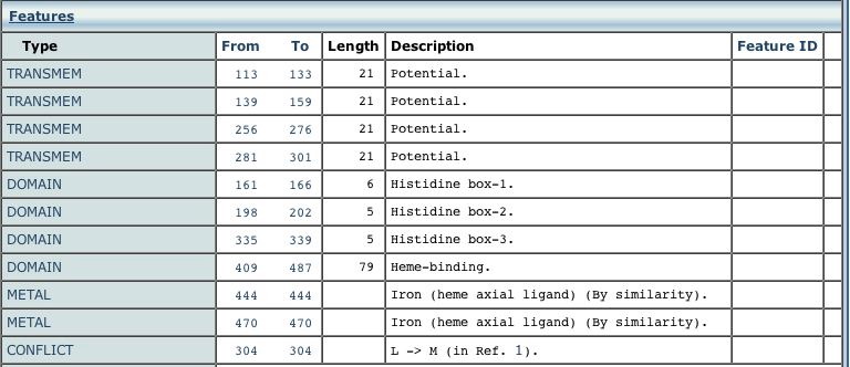
Figure 2. Features of Stearoyl-CoA Desaturase as listed on the UniProt website under information for Stearoyl-CoA. Picture taken from UniProt.
| From Figure 1, it can be seen that Stearoyl-CoA Desaturase has two distinct domains, one being the fatty acid desaturase and the other being the heme-binding domain. Moreover, with the information obtained from the European Bioinformatics Institute's (EBI) Universal Protein Knowledgebase (UniProt), shown in Figure 2, it is evident that these domains can be subdivided into transmembrane domains, Histidine boxes and heme-binding domains. |
(B) YGL050W
Protein Data Bank (PDB): I was unable to find a 3D structure for the protein encoded by YGL050W which makes sense since not much information has been obtained about this protein.
Protein Information Resource (PIR): No information was found on Protein Information Resource about domains in the protein encoded by YGL050W. The picture showed an uncharacterized protein.
Universal Protein Knowledgebase (UniProt): No Information on YGL050W available.
What does it do?
(A) Stearoyl-CoA Desaturase
UniProt: Stearoyl-CoA Desaturase utilizes Iron as a cofactor and catalyzes the following reaction:
Stearoyl-CoA + 2 ferrocytrochrome b(5) + O2 + 2H+ = oleoyl-CoA + 2 ferricytrochrome b(5) + H2O
Munich Information Center for Protein Sequences (MIPS): Stearoyl-CoA Desaturase converts stearoyl-CoA into oleoyl-CoA via electron transport. It is thus, classified as an oxidoreductase.
GO: Stearoyl-CoA Desaturase is involved in mitochondrial inheritance i.e the distribution of mitochondria into daughter cells during mitosis and meiosis, mediated by interactions between the mitochondrion and the cell membrane.
| From the information above, it can be seen that Stearoyl-CoA is important in mediating the changes in the cytoskeleton that occur during cell division, specifically in regards to mitochondrial inheritance. Given that it is responsible for converting saturated fatty acids to unsaturated fatty acids, its role in the dynamic changes taking place in the mitochondrial and cell membrane phospholipids makes sense. |
(B) YGL050W
MIPS: No information on the Biological Process or Molecular Function could be obtained for YGL050W protein.
Where does it live?
(A) Stearoyl-CoA
Gene Ontology (GO): Stearoyl-CoA is located in the Endoplasmic Reticulum.
UniProt: Stearoyl-CoA is an integral membrane protein.
| Since Stearoyl-CoA Desaturase is located in the Endoplasmic Reticulum, its role in mitochondrial inheritance is easy as the ER is a part of the cytoskeleton that offers mechanical support to the cell during these changes. Also, as the new daughter cells form, Stearoyl-CoA is responsible for increasing the degree of unsaturation in the phoshpholipid bilayer of the cell and mitochondrial membranes. |
(B) YGL050W
GO: No information on Cellular Component available.
Who is it related to?
(A) Stearoyl-CoA
MIPS: Stearoyl-CoA has no known paralogs. It, however, does have 19.4% homologs in mammals and 20.8% homologs in bacteria.
| Since Stearoyl-CoA Desaturase has homologs in mammals and bacteria, it is likely that these organisms have similar oxidoreductases that catalyze desaturation reactions as does this enzyme. |
(B) YGL050W
MIPS: YGL050W gene product also does not have any known paralogs. It has 25.6% homologs in humans, 22.0% homologs in archaea and 7.7% homologs in plants.
| From the information obtained from MIPS, it is evident that the YGL050W protein has a significant degree of homology in humans and archaea and some homology in plants. Since the function of this protein has not yet been determined, we can infer that its function is part of a biological process that occurs in humans, plants, archaea as well as yeast. |
Who does it hobnob with?
(A) Stearoyl-CoA
Database of Interacting Proteins (DIP):
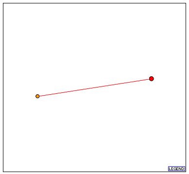
Figure 3. Graph showing the interactions of the protein of interest, stearoyl-CoA (Red dot). The Red dot represents the root protein. It is connected to only one other protein Pmt5p (orange dot). The orange dot represents a 1st shell node, i.e. a protein only one away from the root protein. The red line connecting the two dots represents the connection as deciphered from high-throughput methods but is as yet unverified. Picture obtained from DIP.
GO: Pmt5p is located in the Endoplasmic Reticulum. It is involved in the biological process of O-linked glycosylation.
| From Figure 3, one can see that Stearoyl-CoA is connected to only one other protein called Pmt5p. It appears to be involved in the glycosylation of proteins which is does not really have much connection to lipid metabolism and, more specifically, to the process of desaturation of saturated fatty acids. I could not make a direct connection between these two proteins. |
Kyoto Encylcopedia of Genes and Genomes:
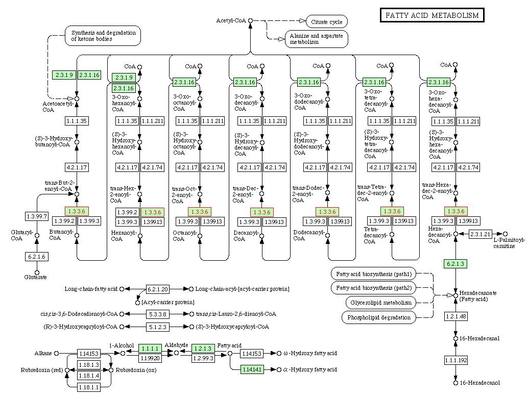
Figure 4. Fatty Acid Metabolism Pathway in S. cerivisiae. The enzyme labeled 1.3.3.6 and highlighted in a red box is acyl-CoA oxidase. The KEGG website shows that this enzyme catalyzes the following reaction: acyl-CoA + O2 = trans-2,3-dehydroacyl-CoA + H2O2. Picture obtained from KEGG.
| Figure 4 illutrates the Fatty Acid Metabolism Pathway in S. cerivisiae. I was unable to locate the exact role Stearoyl-CoA Desaturase plays in this pathway. The enzyme labeled 1.3.3.6, acyl-CoA, is an oxidoreductase, as is Stearoyl-CoA Desaturase. The reaction it catalyzes is very similar to the reaction catalyzed by Stearoyl-CoA desaturase. Thus, although Stearoyl-CoA cannot be found in this pathway, it is likely to be involved in a very similar pathway given the similarity of function of these two enzymes. |
(B) YGL050W
DIP:
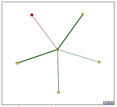
Figure 5. Graph showing the interactions of the protein of interest, YGL050Wp. The red dot represents the protein of interest or root protein, the orange dot represents the 1st shell node, and the yellow dots represent the 2nd shell nodes (i.e. two proteins away from the root protein). The red connecting line represents the connection as deciphered from high-throughput methods but is as yet unverified. The green connecting lines represent connections between proteins that have been verified by one or more computational means. The width of the lines represents the number of experiments performed to establish that connection. Picture obtained from DIP. The orange dot protein in the center is Rad1p. The yellow dot proteins on the periphery are as follows (clockwise from red dot): Rad14p, Msh2p, YAL027W, and Rad10p. Picture obtained from DIP.
GO: Both Rad1p and Rad10p are located in the nucleotide excision repair factor 1 complex. They are both involved in single stranded DNA-specific endodeoxy ribonuclease activity i.e in the catalysis of the hydrolysis of ester linkages in a single stranded DNA molecule. Rad14p also resides in the nucleotide excision repair factor 1 complex and is involved in DNA damage recognition. Msh2p is located in the nuclear chromosome and is involved in DNA and ATP binding during mitotic recombination. The YAL027Wp is located in the nucleus although its molecular function and biological process is unknown.
| From the above information provided by Figure 5 and GO, we can tell that YGL050W is connected to several proteins involved in DNA damage repair. This would imply that YGL050W is also in involved in DNA repair or a related process that occurs during mitosis. If this is the case, then a likely location for this protein would be the nucleus. However, this information contradicts my earlier hypothesis that YGL050W was in some way involved in sporulation and mating when mitosis comes to a halt. |
How can we find it?
From the information available on various websites, the Cellular Component, Biological Process and Molecular Function of the YGL050W protein are not evident. The following experiments may help to determine some of this information: Experiment I. Is YGL050W an essential gene for mating? Procedure: Create an S. cerivisiae YGL050 knockouts using transposons with antibiotic resistant cassettes (to allow for selective growth on antibiotic laden media). Grow the mutant strains for a few hours till log phase growth, and subject the population to alpha-factor at different concentrations. Observe phenotype at various time points for each concentration. Theory: If YGL050W were involved in mating as previously hypothesized, a YGL050W knockout should not be able to reproduce sexually. Sexual reproduction in S. cerivisiae involves the formation of shmoos in response to a certain level of alpha-factor (the mating pheromone) in the surroundings. If YGL050W were involved in mating, shmooing would not be observed in the knockouts. Presence of shmoos would indicate that YGL050W is not essential for this process. Further experiments to examine whether YGL050W mutants can produce appropriate proteins that allow fusion of two yeast cells.
Experiment II. Where is YGL050W found? Procedure: Create S. cerivisiae YGL050W clones with epitope tags. Induce protein and blot cells with fluorescent-tagged antibodies. Examine under microscope for localization of fluorescence. Theory: Clones of YGL050 would produce protein under favorable conditions. YGl050W specific antibodies would bind to the protein and secondary antibodies would bind to the first, causing immunofluorescence. If YGL050W were indeed associated with the Rad proteins, Msh2p and YAL027W as found from the DIP website, it is most likely associated with DNA repair and consequently, located in or near the nucleus. If this is the case, the fluorescent labeling will appear in or near the nucleus. If not, it will appear elsewhere in the cytoplasm. |
Conclusion
The information obtained on Stearoyl-CoA fits in with the previous information obtained about gene expression of OLE1. The information obtained on the YGL050W proteins seems to contradict the gene expression information obtained earlier about the gene YGL050W. While it seems to be expressed in conditions favoring sexual reproduction and sporulation, both of which occur during adverse conditions, the fact that it is functionally connected to proteins involved in DNA repair implies that it may not be connected to sporulation or mating as previously hypothesized. In fact, both sporulation and mating occur when cell division by mitosis cannot due to lack of nutrients, air, etc., while DNA repair occurs specifically during mitotic division. Given the current information, it is difficult to even guess the role of the YGL050W protein, let alone conclusively establish anything. Intensive research on this gene and more concrete data could eventually provide enough information to make an educated guess as to its function and location. |
References
Database of Interacting Proteins. <http://dip.doe-mbi.ucla.edu/dip/Search.cgi?SM=3?>. Accessed November 19th, 2004.
Gene Ontology. <http://www.geneontology.org>. Accessed November 19th, 2004.
Kyoto Encyclopedia of Genes and Genomes. <http://www.genome.jp/kegg/pathway/map/map01100.html>. Accessed November 19th, 2004.
Munich Information Center for Protein Sequences. <http://mips.gsf.de/genre/proj/yeast/index.jsp>. Accessed November 19th, 2004.
Protein Data Bank. <http://www.rcsb.org/pdb/index.html>. Accessed November 19th, 2004.
Protein Information Resource. <http://pir.georgetown.edu/>. Accessed November 19th, 2004.
Universal Protein Knowledgebase. <http://www.ebi.uniprot.org/uniprot-srv/index.do>. Accessed November 19th, 2004.
Back to: