Hematopoietic stem cell gene therapy with a lentiviral vector in x-linked Adrenoleukodystrophy: A summary
This paper was published in the November issue of Science, and it proposes a new treatment for patients that have the cerebral form of Adrenoleukodystrophy (ALD). This paper consists of two sections: preclinical and clinical. The emphasis of this review will be on the clinical portion. Before this study, the main treatment for ALD was a bone marrow transplant. These treatments, apart from being extremely painful and dangerous, require a matching bone marrow donor. As a result, Cartier et al. (2009) “reasoned that hematopoietic stem cell (HSC) gene therapy could be an appropriate therapeutic alternative” for ALD patients that cannot find a bone marrow donor.
This is a list of some words that may be found in this paper. The definitions are courtesy of Horst Ibelgaufts' COPE: Cytokines & Cells Online Pathfinder Encyclopaedia.
Gene Therapy– A therapeutic approach to modify the genome of living cells.
Hematopoietic stem cell (HSC) – These are stem cells that give rise to a variety of cell types (Blood and immune cell types). They are special, because they can renew themselves, differentiate to a specialized cell, and move out of the bone marrow. They differentiate into different cells due to a variety of growth factors and cytokines.
CD34+, CD14+, CD15+, CD3+, and CD19+ – these are HSCs that have been differentiated, and the cells have corresponding cell surface proteins that are recognized by monoclonal antibodies. The antibodies are used to track the location of differentiated HSCs.
Microglial cells– These cells are macrophages (cells that home into areas of inflammation) of the brain and central nervous system that act to fix damaged neurons and plaques.
Engraftment– the process in which stem cells are transplanted to reproduce new cells.
Monocytes– immature cells that, with the correct chemical signal, turn into macrophages.
Lentiviral Vector– A type of retrovirus (Like HIV-1) that infects both diving and non-diving host cells. The virus institutes its own genome into the genome of the host cell through reverse transcription.
In vitro– a procedure that is performed in a controlled environment (not a living organism)
Preclinical
Before testing the lentivirus on the human subjects, it was necessary to make sure that the basic experiment was capable of working. As a result, in vitro studies were performed to determine that the lentiviral vector was capable of inserting the corrected version of the ABCD1 gene (and the viral genome) into a group of host cells. Additionally, studies were performed on ALD protein-deficient mice to determine the ability of the HSCs to replace the diseased mouse microglial cells with new microglial cells that contained functional ALD proteins. Both of these tests were successful, and more importantly, it was determined that the HSCs migrated into the brains of the mice, and then differentiated into microglia that expressed the ALD protein.
Clinical
The subjects in this study were two children, patient 1 and patient 2. Patient 1 was 7.5 years old, and patient 2 was 7 years old. Both patients had the ALD cerebral form, and the ABCD1 gene of patient 1 contained a large deletion of exon 6, while the ABCD1 gene of patient 2 had a missense mutation at position 609 that changed the amino acid from a leucine to a glutamic acid. Additionally, both patients had a progressed state of cerebral demyelination. The absence of the ALD protein in the patients was double checked with immunocytochemistry of the white blood cells. Following this, the patients were injected with a chemical that caused stem cells from the bone marrow to leak into the blood stream. Blood samples were taken, the stem cells were identified, and then taken from the blood. The stem cells were double-checked, and the CD34+ cells were activated with cytokine introduction. The stem cells were then infected with an HIV-1 derived lentiviral vector that was capable of expressing a normal ABCD1 gene. The expression of the gene was under the control of a promoter that could be selectively activated. The transduced cells were then cryopreserved, and the patients underwent a treatment of irradiation to remove the diseased cells, and allow for the maximum engraftment possible. When the procedure was over, the cells were thawed, mixed with cytokines, and engrafted into both patients.
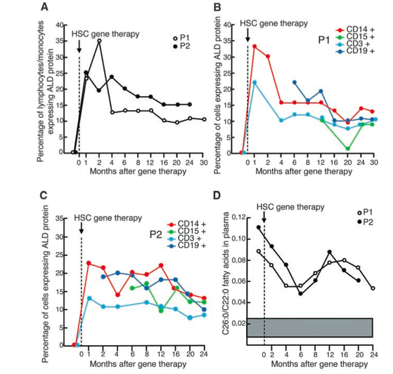
Five days after the modified HSCs were introduced into the patients, it was determined that 50% (patient 1) and 33% (patient 2) of the cells expressed ALD protein. Thirty days after the infusion, the ALD protein was detected in 23% of patient 1’s blood cells, and 25% in patient 2’s blood cells. Every few months, the levels of ALD protein were assessed in each patient, and the ALD protein percentage decreased steadily until about 12 months (Figure 1). From 12 months to 24 months post-infusion, the ALD protein expression of patient 1 decreased from 20% to 18%, and in patient 2 it decreased from 18% to 17%. To determine the levels of the ALD protein, immunofluorescence was used on the bone marrow cells of both patients, and the cells were stained with a CD34+ antibody.
Figure 1: a quantative look at the effectiveness of the HSC gene therapy in both patients over the two year study period.(A) Shows the percentage of monocytes and lymphocytes (immune cells) in both patients over a 30 month period. (B-patient 1)/(C-Patient 2). Depicts the percentage of four different cell types that expressed the ALD protein in relation to the time after the transplant. (D) Portrays the amount of VLCFA in the plasma in relation to the time after the transplant for both patients. The decrease in the VLCFA amount indicates that the HSC method is a potential treatment. Image from Cartier, et al. (2009), permission pending.
The investigators wanted to determine the insertion sites of the lentiviral vector in the stem cells, thus LAM-PCR was performed. To do this, many restriction enzymes were introduced to the DNA (taken from a variety of CD+ bone marrow cells) resulting in smaller, restricted fragments of “cut up” DNA. The restricted DNA fragments were then sorted with Fluorescence Activated Cell Sorting (FACS). The lentiviral insertion points were mapped to points on the human genome, and it was determined that the Lentiviral insertion points were generally in gene coding regions.
Before the gene therapy was performed, an MRI scan of each patient was taken. In patient 1, abnormal and inflamed demyelinated regions of the brain stem, pons, internal capsulae, and periventricular frontal white matter were found. In patient 2, there were more extensive demyelinating lesions throughout the corpus callosum, white matter of the parietoccipital lobes, and the auditory tract. Throughout the experiment, some of the demyelinating lesions increased in size, however they eventually reached a stable point. Additionally, parts of the neuronal lesions decreased over time indicating a reversal in the demyelination process (Figure 2). Lastly, the very long chain fatty acids (VLCFA) decreased by 39% in patient 1 (24 months after infusion) and 38% in patient 2 (20 months after infusion). This result was not expected due to the seemingly low percentage of the ALD protein in the blood cells (Figure 2).
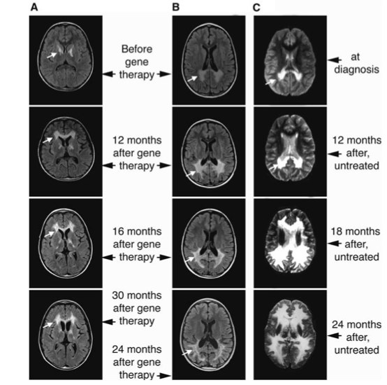
Figure 2: A comparison of brain MRI images of ALD patient 1(A) and ALD patient 2(B) in relation to a person with ALD that is not treated (C). The images are taken before and after the HSC gene therapy in patient 1 and patient 2. It appears that the demylenating lesions of the HSC treated patients are stabilized and/or or slightly retreated. Additionally, the HSC treated patients have dramatically less neuronal lesions than an untreated ALD patient. Image from Cartier, et al. (2009), permission pending.
The initial progression of the lesions, and then their stabilization/decrease is a common result to what is seen in patients that are treated with bone marrow transplants. Patients that are subjected to bone marrow transplants generally observe no changes in the demyelination process for many years after the operation. Thus, this study is hopeful that the Hematopoietic stem cell (HSC) gene therapy will have similar results in the long run. In conclusion, the investigators found that the lentiviral vector use in Hematopoietic stem cell gene therapy may be a potentially successful treatment for patients with ALD that cannot find a bone marrow donor. Additionally, they believe that the methods used in this paper may provide a method for gene therapy in patients with other CNS diseases.
For more information on lentiviral vectors and gene therapy, click here.
References:
Horst Ibelgauft. Cytokines & Cells Online Pathfinder Encyclopedia. Summer 2010 [cited 2011 Jan 28]. Available from: http://www.copewithcytokines.de/cope.cgi?key=PBMC
Nathalie Cartier, Alima Hacein-Bey-Abina, Cynthia C. Bartholomae, Gabor Veres, Manfred Schmidt, Ina Kutschera, Michel Vidaud, Ulrich Abel, Liliane Dal-Cortivo, Laure Caccavelli, et al. Hematopoietic Stem Cell Gene Therapy with a Lentiviral Vector in X-Linked Adrenoleukodystrophy Signal processing in single cells. Science [Internet]. 2009 November 6[cited 2011 Jan 17]; 326(5954): 818-822 . Available from: http://www.sciencemag.org/content/326/5954/818.short