Molecular Tool: Ligation
This web page was produced as an assignment for an undergraduate course at Davidson College.
General
Information
Ligases are
enzymes that seal breaks in the phosphate-sugar backbone of DNA and RNA. There
are two main classes of DNA ligases, those that use NAD+ as a cofactor (only in
bacteria), and those that use ATP as a cofactor (eukaryotes and viruses) (Kahn,
2003). Mammalian cells have four types of DNA ligase, which together accomplish
three main functions: joining Okazaki fragments, sealing repairs, and sealing
recombination fragments (Purves et al., 2001).
DNA ligase I: connects Okazaki fragments of the lagging strand in
DNA replication, and can also seal some repair and recombination fragments (Wei
et al., 1995).
DNA ligase II: an alternatively spliced form of DNA ligase III that
is only expressed in non-dividing cells.
DNA ligase III: works with protein XRCC1, which is a DNA repair
protein. DNA ligase III is a primary agent in sealing base excision-repairs and
recombination fragments.
DNA ligase IV: works with protein XRCC4, which is another DNA repair protein. DNA ligase IV is also important in sealing base excision-repairs and recombination fragments, especially during development (Schär et al., 1997).
Mechanism
The following diagrams and
descriptions are of the three main steps of ligation among all types of DNA
ligases. Notice how ligase follows the general properties of enzymes, solely
providing a docking area for the reaction between the AMP and DNA.
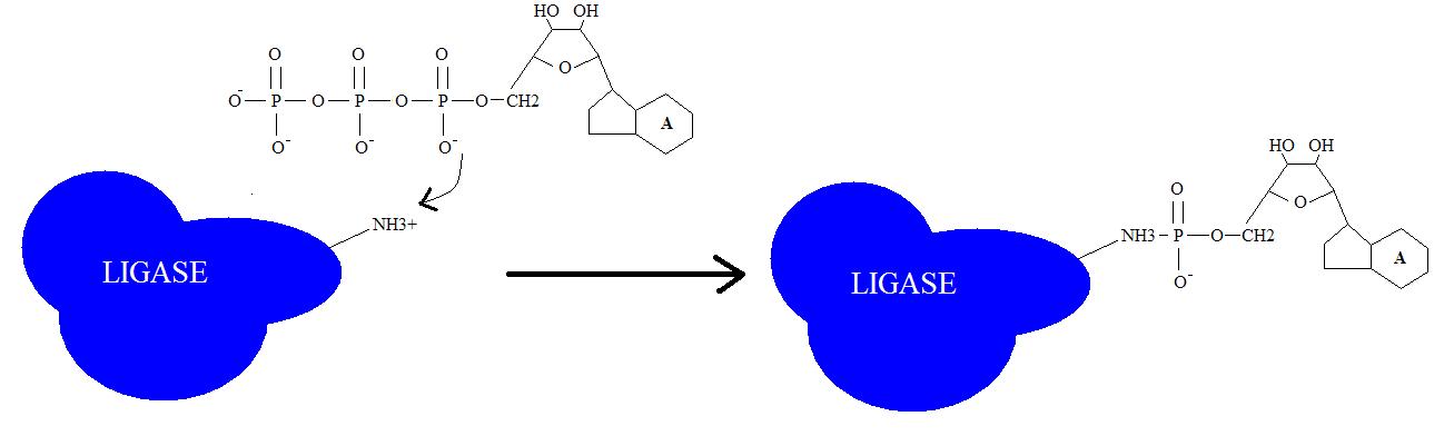
1. Adenylation of DNA ligase:
The side chain of lysine 34 in ligase forms a bond with ATP, where ATP kicks off two phosphate groups to become an AMP-ligase complex (Kahn, 2003).
|
Figure 1. Adenylation of DNA ligase by ATP. |
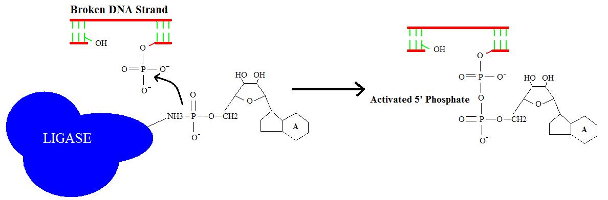
2. Activation of 5’
phosphate:
The monophosphate of the
AMP-ligase complex forms a bond with the 5’ phosphate of the broken strand.
This bonding activates the 5’ phosphate group for the next step.
|
Figure 2. Activation of 5’ phosphate mediated by AMP-ligase complex. |
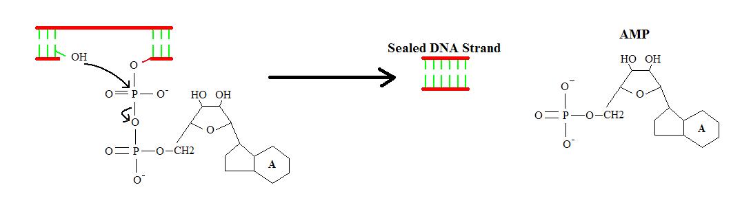
3. Displacement of AMP connects the broken strand:
Now that the 5’ phosphate has
been activated by the AMP-ligase complex, the 3’ hydroxyl group attacks the 5’
phosphate and forms a new bond, releasing the AMP. Ligase is crucial for
holding the complex together in the necessary orientation.
|
Figure 3. Displacement of AMP connects broken strand with catalytic help by ligase (not shown). |
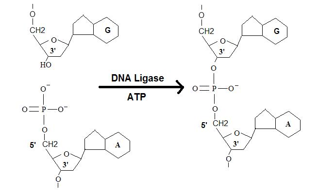
The Overall Reaction:
|
Figure 4. Overall reaction of the ligation of a broken DNA strand to form a new phosphodiester bond between the adjacent phosphate and sugar. |
Using Ligation as a Molecular Tool
|
Figure 5. Chime image of ATP-dependent DNA ligase from bacteriophage T7 complex with ATP. Notice the location of lysine 34 within ligase that will eventually form a covalent bond with AMP. Click Here for Source: PDB |
T7 DNA ligase is approximately half the size of human DNA ligase, having a molecular weight of 41 KdA (Kahn, 2003). However, T7 DNA ligase is a versatile tool for molecular biologists because it shares common structure with a wide spectrum of ligases from different species. As mentioned before, ligases seal breaks in the phosphate-sugar backbone of adjacent nucleotides in DNA or RNA. In creating expression vectors, and many synthetic DNA fragments, ligases are essential in linking the pieces of the puzzle. The most efficient way for a ligase to link DNA fragments is to connect two sticky ends together from a restriction digest. The hydrogen bonds between the complementary bases aid ligase in holding the two ends together while the phosphodiester bond is formed. Although ligases are capable of connecting two blunt ended DNA fragments together, the reaction is much slower and prone to greater error. Blunt end ligation should have controls to demonstrate that the proper fragments were ligased because the specificity decreases from sticky end ligation. Ligations are relatively fast reactions at 16o C, usually requiring 5-30 minutes to completely ligate a sample (Kahn, 2003). One limitation to ligation is that it requires a 5’ phosphate on the DNA fragment to work at all, however T4 polynucleotide kinase and ATP can usually phosphorylate the 5’ carbon in the broken DNA strand (Kahn, 2003).
References
Purves, W., Sadava, D., Orians, G., Heller, H. Life:
The Science of Biology. Sinauer Associates, Inc., 2001.
Kahn, J. "DNA Ligase." University of
Maryland Deparment of Biochemistry. Visited on 13 Feb. 2003. http://adnadn.umd.edu/biochem/kahn/molmachines/replication/DNA%20Ligase.htm
Schär, P., Herrmann, G., Daly, G., and Lindahl, T.
(1997). A newly identified DNA ligase of Saccharomyces cerevisiae involved in
RAD52-independent repair of DNA double-strand breaks. Genes Dev. 11, 1912-1924.
(Site visited on 13 Feb. 2003: http://www.imr.unizh.ch/research/schaer/ligase.html)
Wei, Y.-F., Robins, P., Carter, K., Caldecott, K.,
Pappin, D. J. C., Yu, G.-L., Wang, R.-P., Shell, B. K., Nash, R. A., Schär, P.,
Barnes, D. E., Haseltine, W. A., and Lindahl, T. (1995). Molecular cloning and
expression of human cDNAs encoding a novel DNA ligase IV and DNA ligase III, an
enzyme active in DNA repair and recombination. Mol. Cell. Biol. 15, 3206-3216.
(Site visited on 13 Feb. 2003: http://www.imr.unizh.ch/research/schaer/ligase.html)
![]()
![]()
© Copyright 2002 Department
of Biology, Davidson College, Davidson, NC 28036
Send comments, questions, and suggestions to: drweber@davidson.edu