Hemoglobin
(JMOL Here)
What is hemoglobin?
Found in red blood cells, hemoglobin are globular proteins that ferry oxygen (O2) molecules and carbon dioxide (CO2) molecules throughout the body. Each hemoglobin protein structure consists of four polypeptide subunits, which are held together by ionic bonds, hydrogen bonds, hydrophobic interactions, and van der Waals forces, as well as four heme pigments, one in each of the subunits (Sadava et al., 2008). These heme groups contain positively-charged iron (Fe2+) molecules which can reversibly bind to oxygen molecules and transport them to various areas of the body (Sadava et al., 2008). As the heme groups bind or release their oxygen loads, the overall hemoglobin undergoes conformational changes which alters their affinity for oxygen (Sadava et al., 2008).
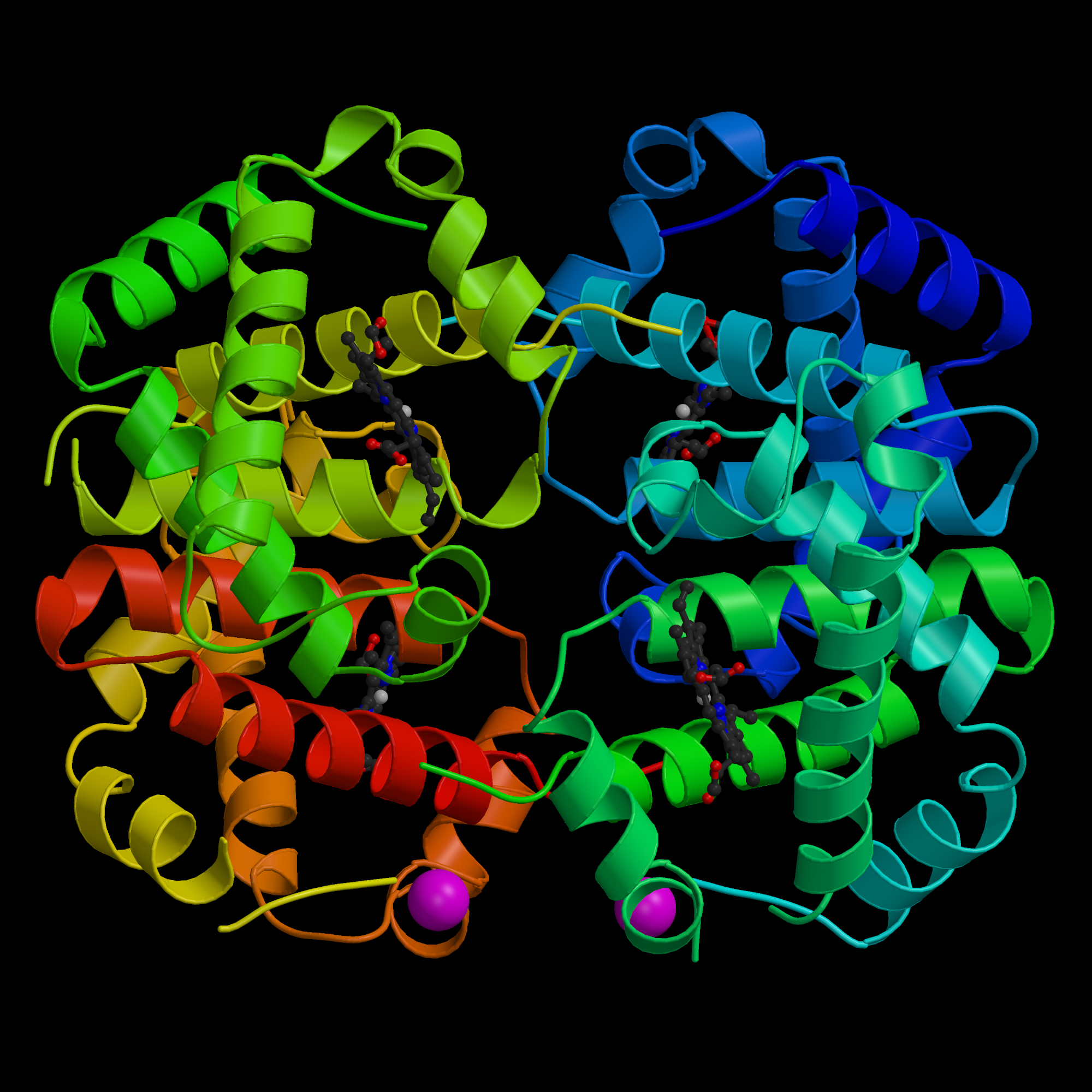
Figure 1. Structure of hemoglobin. There are four subunits as shown
by the various colors. The heme groups are shown in predominantly gray.
http://ucsdnews.ucsd.edu/newsrel/supercomputer/04-08ProteinDataBank.asp.
Permission pending.
Structure and Function
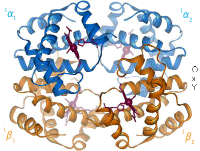
Hemoglobin
tetramers are comprised of the four subunits, two α-globin chains and two β-globin
chains all of which take the form of alpha helices (Sadava et al., 2008). Found in each chain is a non-protein
heme group, which is an assembly of cyclic ring structures surrounding an iron
ion that is tethered by nitrogen atoms (Perutz, 1990). The heme group, which is
typically hidden within the various subunits, is covalently bound to yet a
different nitrogen atom that belongs to a nearby histidine group (Perutz, 1990).
This histidine chain, combined with other hydrophobic interactions, stabilize
the heme group within each subunit. Oxygen molecules bind to the side of the
iron ion that is opposite of the proximal histidine. Located near this opposite
side is a different histidine chain, which serves two important function even
though it is not directly bound to the heme group (Perultz, 1990).

Figure 2. Heme molecule. Four Nitrogen molecules interact with
Iron atom surrounded in cyclic ring structures.
From http://en.wikipedia.org/wiki/Hemoglobin. Permission pending.
Because
histidine is itself positively charged, its close proximity to the negatively
charged iron ion prevents the iron ion from becoming too oxidized, which would
inhibit the binding of oxygen molecules. This is critical to the hemoglobin’s
job of oxygen transport, since oxygen can bind to Fe2+, but not Fe3+
(Boyer, 2006). Also, the size, shape, and location of this distal histidine
chain limits the amount of CO2 molecules that will bind to the heme group
(Boyer, 2006). Because the heme group has a greater natural affinity for carbon
monoxide than for oxygen, the lack of this distal histidine chain would allow
heme groups to bind significantly more to carbon waste than to oxygen,
preventing hemoglobin proteins from providing cells with the necessary oxygen
molecules for metabolic activities (Boyer, 2006).
After an oxygen molecule binds to one of the heme groups of any subunit, other subunits undergo conformation changes exposing their own heme groups, thus giving the entire hemoglobin structure greater oxygen affinity (Perutz, 1990). The bond between oxygen the oxygen atom and the iron ion pulls the iron molecule closer to its heme group, which then pulls the proximal distal histidine chain backwards into the hemoglobin molecule (Perutz, 1990). This pull creates a strain on the other subunits, breaking ionic bonds in such a way that reveals their obscured individual heme groups. This positive cooperation allows binding at one subunit to increase the binding affinity at other subunits (Sadava et al., 2008). A visual representation of this pulling can be found here.
Works Cited
Boyer R, et
al. 2006. Myglobin & Hemoglobin. http://www3.interscience.wiley.com:8100/legacy/college/boyer/0471661791/structure/HbMb/hbmb.htm.
February 2010.
Neer E,
Konisberg W, and Guidoti G. The Interactions Between α and β chains of Human
Hemoglobins. Journal of Biological
Chemistry, 1967; 243: 1971-1978.
Perutz M.F. Mechanisms Regulating the Reactions of
Human Hemoglobin with Oxygen and Carbon Monoxide. Annual Review of Physiology,
1990; 52: 1-26.
Sadava D,
Heller CH, Orians GH, Purves WK and Hillis DM. Life: The Science of
Biology 8th Edition. Massachusetts and Virginia: Sinauer Associates Inc. and W.H.
Freeman and Company, 2008. Print.
Please send suggestions, questions, or comments to tahua@davidson.edu