From PDB database at http://www.rcsb.org/pdb/cgi/explore.cgi?pid=225481005252650&pdbId=1D8B
UNANNOTATED YEAST PROTEIN-YJU3
SUMMARY OF INFO FROM THE PREVIOUS TWO WEB SITES
Analysis of YJU3s conserved domains suggest that
it is a lysophospholipase. Lysophospholipases remove the second fatty
acid from a phospholipid after the first fatty acid is removed by a phospholipase
(Cox, 2000). Microarray data indicate that the YJU3 gene is strongly
induced during a diauxic shift (SGD,
2001). Specifically YJU3 is not induced until glucose levels nearly
reach zero. The data suggest that YJU3 may be involved in the breakdown
of phospholipids to produce energy when glucose is not available.
NEW EXPERIMENTS
The Yale Gerstein Lab used protein macroarrays to
discover what proteins bound phospholipids (http://spine.mbb.yale.edu/protein_chips/).
YJU3 was not found to bind to any of the phospholipids tested in this experiment.
Considering YJU3 has highly conserved domains with lysophospholipases,
one would have expected that YJU3 would interact with phospholipids.
However, since lysophospholipases cleave the second fatty acid from phospholipids
only after the first fatty acid is removed, it would be interesting to
redo this experiment using phospholipids that have had the first fatty
acid removed. YJU3 may be found to interact with these modified phospholipids.
In another experiment, Snyder
and his team (2000) created a unique transposon that allowed them to
observe when a gene was expressed using a lacZ gene, and where the gene
was expressed using an epitope tag. Among the strains Snyders lab
produced, three strains containing the transposon in the YJU3 gene were
created. The targeted YJU3 genes showed light expression during sporulation
and vegetative growth. No significant phenotype disruptions
were observed under the environmental conditions tested by Snyders lab.
There was also no staining above background levels when grown in YPD medium.
http://ygac.med.yale.edu/triples/basic_search.asp
Considering the microarray data, it may prove advantageous
to observe YJU3 transposon mutants undergoing a diauxic shift. The
YJU3 transposon mutants may exhibit abnormal growth and phenotypes when
the cell is exposed to these conditions if the YJU3 protein is responsible
for providing energy from the breakdown of phospholipids when glucose is
depleted. If YJU3 mutants respond under these conditions, the phenotypes
that result may help elucidate the functions of the YJU3 protein.
Also the localization of the YJU3 protein when it is active may be discovered.
Using a technique developed by
Brian Chaits lab (1999), protein levels of the YJU3 protein between
yeast undergoing a diauxic shift and yeast growing in normal conditions
could be compared. For example yeast undergoing a diauxic shift
could be grown in a media containing heavy nitrogen and yeast growing under
normal conditions could be grown in a media containing light nitrogen.
Then the levels of the YJU3 protein could be evaluated using mass spectroscopy.
If YJU3 is actively translated during a diauxic shift then there should
be a significant difference in the amount of YJU3 protein observed between
these two conditions.
Brian Chaits lab at the Rockefeller University
in NYC (1999) also used stable isotopes to observe differences in phosphorylation
of a protein between two conditions. This method could be used to discover
if the YJU3 protein is phophorylated during a diauxic shift. If YJU3
is regulated by phosphorylation then there should be a reciprocal change
in the amount of YJU3 protein that is phosphorylated during a diauxic
shift.
If the YJU3 protein is regulated by phosphorylation
then it would be useful to use a protein chip to discover what kinase does
the phosphorylation using the technique developed by Mike Snyders lab
(2000). Specifically the YJU3 gene could be spotted in all of the
wells on a chip. Then a different kinase plus radioactive ATP
could be added to each well. A resulting dark spot would suggest
that the kinase may be involved in the regulation of the YJU3 protein through
phosphorylation. http://bioinfo.mbb.yale.edu/genome/yeast/chip/
NO USEFUL INFO FOUND YET
DIP
No information about YJU3 was found.
http://dip.doe-mbi.ucla.edu/dip/Search_DIP.cgi
Two Hybrid
YJU3 was not in the Fields lab database for yeast two hybrid experiments.
http://depts.washington.edu/sfields/yplm/data/index.html
Path Calling Interaction Database
A search for YJU3 produced no protein interactions.
http://portal.curagen.com/extpc/com.curagen.portal.servlet.PortalYeastList
ANNOTATED YEAST PROTEIN-SGS1
SUMMARY OF INFO FROM PREVIOUS WEB SITES
SGS1 is a DNA helicase involved in transcription
of rRNA and replication (SGD,
2001). SGS1 is thought to suppress illegitimate recombination and interact
with RNA polymerase II during rRNA transcription (SGD, 2001). SGS1
interacts with Top3, Top2, (YPD
protein report, 2001) and srs1 proteins (Lee, 1999) to perform these
functions. SGS1 is also thought to play a role in cell cycle checkpoints
(Frei, 2000; McVey, 2001). SGS 1 is interesting because it is a homologue
to the gene that causes Werners and Blooms syndrome in humans (YPD protein
report, 2001). Blooms syndrome results in increased incidences of
cancer, genomic instability, and growth retardation (Watt, 1996).
Werners syndrome results in premature aging (Guarente, 1997). SGS1
in yeast is being used a model for these to diseases.
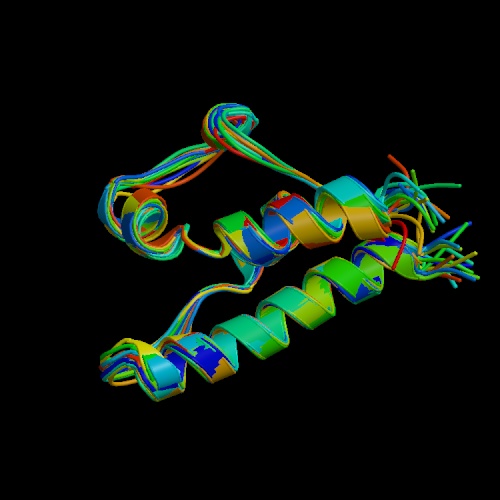
The Hrdc domain of the SGS1 protein has been crystalized.
From PDB database at http://www.rcsb.org/pdb/cgi/explore.cgi?pid=225481005252650&pdbId=1D8B
NEW INFO
Protein Interaction Map
In the protein interaction map, SGS1 interacts with
TOP3 and AUT7 and AUT7 in turn interacts with TOP2 (Schwikowski, 2000).
AUT7 attaches autophagosomes to microtubules (SGD,
2001). TOP2 is involved in meiotic recombination and DNA elongation
(SGD,
2001). TOP3 is a topoisomerase that works with the SGS1 helicase
to regulate meiotic recombination (SGD,
2001).
In the main protein interaction map, SGS1 and TOP3 are said to have
similar roles but different locations and SGS1 and AUT 7 are said to have
the same location but different functions. In the other protein interaction
maps SGS1 is classified as being involved in meiosis, aging, and RNA processing
and modification. AUT7 is grouped with proteins involved with protein
degradation, vesicular transport, RNA turnover, and meiosis. TOP3
is categorized as being involved in chromosome structure, meiosis, and
recombination.
Protein interaction maps can be found at this web site: http://depts.washington.edu/sfields/yplm/data/index.html
NEW EXPERIMENTS
Since SGS1 is involved in meiosis, transcription,
and cell cycle check points, the SGS1 protein is probably regulated in
some fashion. To discover whether phosphorylation plays a role in
the regulation of SGS1, the SGS1 protein could be analyzed using a protein
chip technique developed by Snyders lab and a stable isotope method developed
by Brian Chiats lab. In Snyders method SGS1 protein would be spotted
in every well of a protein chip. Then a different kinase and radioactive
ATP would be added to each well. If the kinase phosphorylated SGS1
then a dark spot would appearl. In the stable isotope method developed
by Chiats lab, cells could be stalled at different stages in growth in
a media containing normal nitrogen. When the cells were allowed to
proceed with normal growth heavy nitrogen could be introduced to the medium.
Observing the rations of heavy to light nitrogen may elucidate the time
and stage that the SGS1 protein is being phosphorylated.
There is not a lot of evidence supporting
the AUT7 SGS1 interaction predicted by the protein interaction map developed
by Schwikowski (2000). AUT7 and SGS1 are predicted to be a link between
Top3 and Top2 proteins. This suggests that the AUT7 SGS1 interaction
takes place during meiosis. It would be interesting to observe the
protein levels of these two proteins at different stages in a cell's life
using the stable isotope method developed by Brian Chiats lab (1999).
It would also be interesting to see if the two proteins are phosphorylated
at the same times or reciprocally phosphorylated using Brian Chiats stable
isotope method (1999). Any correlation between the two proteins may
help elucidate when the two proteins are working together.
NO USEFUL INFO FOUND YET
Two Hybrid
Stan Fields Lab has no data regarding SGS1.
http://depts.washington.edu/sfields/yplm/data/index.html
Since SGS1 interacts with DNA it is a good candidate for the yeast
two hybrid approach.
Path Calling Interaction Database
SGS1 was not found to interact with any proteins.
http://portal.curagen.com/extpc/com.curagen.portal.servlet.PortalYeastList
DIP database
The DIP database shows SGS1 interacting with Top3.
http://dip.doe-mbi.ucla.edu/dip/Search_DIP.cgi
CHAITS LAB
Oda, Y., K. Huang, F.R. Cross, D. Cowburn, and Brian
T. Chait. 1999. PNAS. Vol. 96:
6591-6596.
Cox, Michael and David Nelson. 2000.Lehninger Principles of Biochemistry.
Worth Publishers.
New York. 363-384.
FIELDS LAB
http://depts.washington.edu/sfields/
YEAST TWO HYBRID
Uetz P et al. (2000) A comprehensive
analysis of protein-protein interactions in
Saccharomyces cerevisiae [PDF; 480 KB!]. Nature, Feb 10, 403 (6770): 623-627.
PROTIEN INTERACTION MAPS
Schwikowski B et al. (2000)
A network of protein interactions in yeast. Nature Biotechnology
Dec. 2000, in press.
http://depts.washington.edu/sfields/yplm/data/index.html
Frei, Christian and Susan M. Gasser. January 2000. The yeast Sgs1p helicase
acts upstream of
Rad53p in the DNA replication checkpoint and colocalizes
with Rad53p in S-phase-specific
foci. Genes and Dev. 14(1): 81-96.http://www.genesdev.org/cgi/content/full/14/1/81
Guarente, Leonard. October 1997. Link between aging and the nucleolus.
Genes and Dev. 11(19):
2449-2455.
http://www.genesdev.org/cgi/content/full/11/19/2449
Lee, S. K. , Johnson, R. E. , Yu, S. L. , Prakash, L. & Prakash,
S. 1999. Requirement of Yeast
SGS1 and SRS2 genes for replication and transcription.
Science 286: 2339-2342.
http://www.sciencemag.org/cgi/content/full/286/5448/2339?ijkey=bWVP.CI6.mh6A
McVey, M. , Kaeberlein, M. , Tissenbaum, H. A. & Guarente, L. 2001.
The short life span of
Saccharomyces servisiae sgs1 and srs2 mutants is
a composite of normal aging processes and
mitotic arrest due to defective recombination. Genetics
157: 1531-1542.
http://www.genetics.org/cgi/content/full/157/4/1531
PATH CALLING INTERACTION DATABASE
02/10/00 - A collaboration between Stanley Fields'
Lab at the University of Washington, Dept.
of Genetics & Howard
Hughes Medical Institute and CuraGen Corporation has completed
a genome-wide analysis of
the protein-coding genes of the yeast genome. This analysis
identifies proteins
which are likely to form stable complexes with other proteins.
http://portal.curagen.com/extpc/com.curagen.portal.servlet.PortalYeastList
http://ygac.med.yale.edu/default.stm
TRIPLES DATA BASE
http://ygac.med.yale.edu/triples/triples.htm
Kumar,
A., Cheung, K.-H., Ross-Macdonald, P., Coelho, P.S.R., Miller, P., and
Snyder,
M. (2000). TRIPLES: a Database of Gene Function in S. cerevisiae. Nucleic
Acids Res.
28, 81-84. (Full-text in PDF reproduced with permission from NAR Online
http://www.oup.co.uk/nar
)
Ross-Macdonald,
P., Coelho, P.S.R., Roemer, T., Agarwal, S., Kumar, A., Jansen,
R., Cheung, K.-H., Sheehan, A., Symoniatis, D., Umansky, L., Heidtman,
M., Nelson,
K., Iwasaki, H., Hager, K., Gerstein, M., Miller, P., Roeder, G.S., and
Snyder, M.
(1999). Large-scale analysis of the yeast genome by transposon tagging
gene
disruption.Nature 402, 413-418.
PROTEIN-KINASE INTERACTION
http://bioinfo.mbb.yale.edu/genome/yeast/chip/
H
Zhu, J Klemic, S Chang, P Bertone, A Casamayor, K Klemic, D Smith, M Gerstein,
M
Reed,
& M Snyder (2000). Analysis of yeast protein kinases using protein
chips. Nature
Genetics 26: 283-289.
SGD database. 2001.Stanford.
http://genome-www.stanford.edu/Saccharomyces/
Watt, Paul M. and Ian D. Hickson. 1996. Failure to unwind causes cancer.
Current Biology.
6:265-267. http://journals.bmn.com/journals/list/browse?uid=JCUB.bb6308&rendertype=text
YPD database. 2001. Proteome, Inc.
http://www.proteome.com/databases/index.html
Yale Gernstien Lab
http://spine.mbb.yale.edu/protein_chips/
Send comments to: lirobinson@davidson.edu