Photo from Genetics Science Learning Center, University of Utah
Visualization of Chromosomes
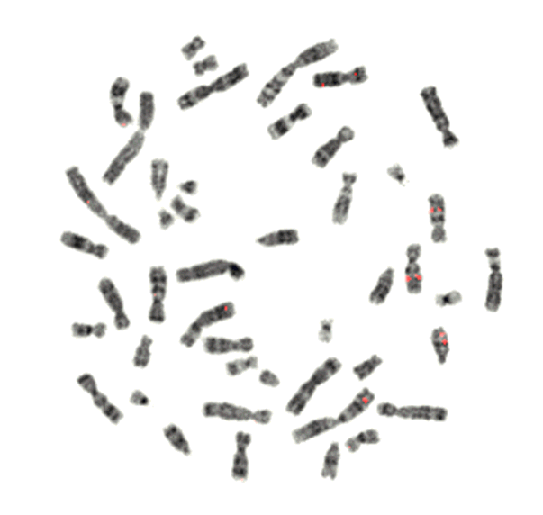
BACKGROUND
As we will discuss in lecture, chromosomes become highly condensed during the initial
stages of mitosis and meiosis. This chromosomal condensation results in the
chromosomes becoming visible with basic light microscopy. At this stage, then,
researchers easily can study the chromosomal structure and determine the karyotype
of the cell. Such studies typically are used to screen for gross chromosomal
abnormalities such as aneuploidy, translocations, and inversions. Various chromosomal
staining techniques also have been developed which allow researchers to identify
specific landmarks within the chromosomes. In this exercise, we will isolate,
stain, and examine the chromosomes from a mouse fibroblast cell line.
A
Photo from Genetics Science Learning Center,
University of Utah
GOALS
Learn basic cell culture techniques
Refresh basic microscopy techniques
Investigate chromosome structure
Think about karyotypes
(photo from Genetics Science Learning Center, University of Utah)
MATERIALS
Human HeLa cells
Colchicine
Methylene blue
Microscope slides
Light microscopes. Need
a refresher course on microscopes?
PROTOCOL
Prior to the laboratory period, human HeLa cells were treated with colchicine and then resuspended in ethanol.
Preparation of chromosome spreads:
1. Gently vortex the tube containing the HeLa cells.
2. Remove 50ul of cells.
3. Allow a drops of the cells to fall
onto a slide at a 45 degree angle from a distance of 2-3 feet.
12. Allow the slide to air dry.
Staining of chromosomes:
1. Add 1 drop of methylene blue to the area containing the cells.
2. Incubate for 10 minutes at room temperature.
3. Aspirate the excess methylene blue.
4. Allow slide to air dry.
5. Add a drop of mounting medium to the slide and apply a cover slip.
6. View chromosomes with light microscopes.
Terms
Colchicine
Metaphase
Diploid
Polyploid
Aneuploid