Simple Columnar Epithelium
Structure
 |
Katy Sims |
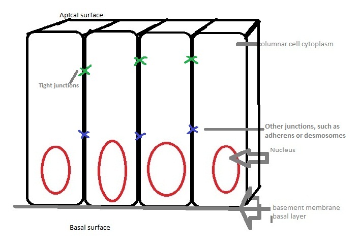
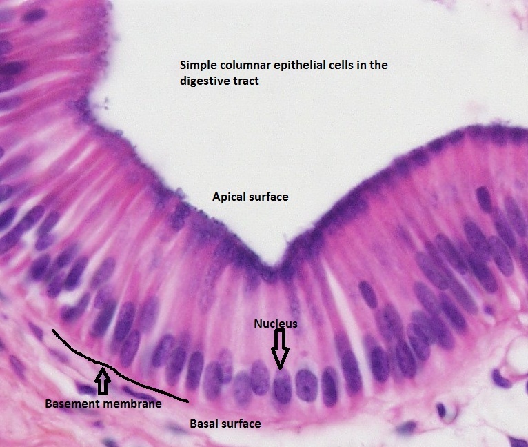
| Simple columnar epithelial cells are longer than they are wide. Characteristically, their nucleui are found at the base of the cell. The cells are connected by tight junctions. The cells receive nutrients through the basement membrane, which separates the cells from the capillary basal layer. |
Location
 |
adapted from dreamstime.com
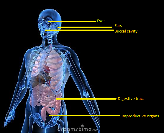
Simple columnar epithelial cells are some of the most prolific cells in the body, mainly because they can fulfill so many functions. They are found throughout the body's organ system, including the digestive tract and the female reproductive system. They are found in the respiratory system, including the nasal passage. Because epithelium can be innervated, simple columnar epithelial cells can also serve a sensory function: simple columnar epithelium line the ears and buccal cavity and are found in the eye. Epithelia is found in so many places due to its variable permeability, polarized gradient (INTERNAL LINK TO POLARITY), and barrier function (INSERT LINK TO TIGHT JUNCTIONS).
Function
 |
| The main function of simple columnar epithelial cells are protection. For example, the epithelium in the stomach and digestive tract provides an impermeable barrier against any bacteria that could be ingested but is permeable to any necessary ions. This function is especially important in the colon. Simple columnar epithelial cells can specialize to secret mucus that coats and protects the surrounding surface from damage(INSERT LINK). Because the epithelium can be innervated, simple columnar epithelium is also specialized to provide sensory input. These cells are found in the cornea, inner ear, and nose. Finally, simple columnar epithelium are very good at absorping and transporting nutrients from locations like the small intestine. See our home page for more descriptions of epithelial functions (INSERT LINK). |
Sources
Ackerley, S. K. Developmental Biology Online. “Epithelial cells”. 09 Dec 2005. 04 Sept 2010. http://www.uoguelph.ca/zoology/devobio/210labs/epithelial1.htmlhttp://www.uoguelph.ca/zoology/devobio/210labs/epithelial1.html
King, D.. Histology at SIU SOM. “Epithelium Study Guide”. 8 Feb 2010. 5 Sept 2010. http://www.siumed.edu/~dking2/intro/epith.html
Sadava, D., Heller, H. C., Orians, G. H., Purves, W. K., & Hillis, D. M. (2008). Life: The Science of Biology (8th ed.). U.S.A.: Sinauer Associates, Inc. (Original work published 2006)
Wann, David. "David Wann's Web Page 1." Carl Albert State University. 12 Jan 2010. 02 Oct 2010. http://www.carlalbert.edu/dwann/tissue_images/simple%20columnar%20epithelium.jpg