Fall 1997 Biology 111 Exam #2 - Genetics through linkage
Dr. Campbell's course
There is no time limit on this test, though I have tried to design one that you should be able to complete within 2.5 hours, except for typing. You are not allowed to use your notes, old tests, or any books, nor are you allowed to discuss the test with anyone until all exams are turned in at 10:30 am on Monday October 20. EXAMS ARE DUE AT CLASS TIME ON MONDAY OCTOBER 20. You may use a calculator and/or ruler. The answers to the questions must be typed on a separate sheet of paper unless the question specifically says to write the answer in the space provided. If you do not write your answers on the appropriate pages, I may not find them unless you have indicated where the answers are.
Please do not write or type your name on any page other than this cover page. Staple all your pages (INCLUDING THE TEST PAGES) together when finished with the exam.
Name (please print):
Write out the full pledge and sign:
How long did this exam take you to complete (excluding typing)?
Lab Questions:
3 pts.
1) What is the total magnification of the microscope when the light is set
for 50%, the condenser aperture is opened to 25%, the ocular says 16X, the
objective lens says 40X, and there are only 20 microliters of cells on the
microscope slide?
16 x 40 = 640
5 pts.
2) One particularly keen student volunteered to come to an extra lab
session over fall break. During this time, she made the following observations
after a mating of Chlamydomonas gametes:
27 cells with four flagella and 8 cells with two flagella
Determine the mating efficiency if the formula is:
2(no. of zygotes) divided by [2(no. of zygotes) + (no. of gametes]
2(27) / [2(27) + 8] = .87 or 87%
2 pts.
3) How long is a typical Chlamydomonas flagellum when it is full
length?
2 - 3 microns (µm)
Lecture Questions:
3 pts.

4) Is the disease trait above dominant, codominant, or recessive? To receive
full credit, you must explain how you have reached your conclusion.
Recessive. Since the parents do not display the
phenotype and all the children do, both parents must have been carriers
for the disease. Unfortunately, they were very unlucky to have all four
children be homozygous recessive.
6 pts.
5) a) Is the disease trait above dominant, codominant, or recessive? To
receive full credit, you must explain how you have reached your conclusion.
Dominant. Since both parents have the disease but
some children do not have it, the parents cannot be homozygous recessive.
If they were homozygous recessive, then all of their children would have
to have the disease too. Instead, the parents are heterozygous with one
dominant disease allele and one recessive wild-type allele.
b) Provide the genotypes (as best you can) of everyone in this family.
(D = disease allele; d = wild-type allele
1 = Dd, 2 = Dd, 3 = DD or Dd, 4-6 all = dd.
Do not use any information in question #6 to influence your answer
for question #5.
12 pts.
6) Use the pedigree with numbers and now make the assumption that a person
cannot reproduce if he or she is a homozygote with the disease alleles.
For couple 1 x 2: 1 = Dd and 2 = Dd
a) What are the odds that their next child will have the disease?
3/4
b) What were the odds that they would have three children without the
disease and one with?
1/4 x 1/4 x 1/4 x 3/4 = 3 /256
+ 1/4 x 1/4 x 3/4 x 1/4 = 3 /256
+ 1/4 x 3/4 x 1/4 x 1/4 = 3 /256
+ 3/4 x 1/4 x 1/4 x 1/4 = 3 /256
total odds = 12/256 = 3/64
c) What are the odds of having the first child with the disease and the
next three without the disease?
3/4 x 1/4 x 1/4 x 1/4 = 3/256
d) What are the odds that there next child will be a boy without the
disease?
1/2 x 1/4 = 1/8
4 pts.
7) During which phase, or phases, of mitosis and meiosis do homologous chromosomes
separate?
mitosis - homologous chromosomes are never paired;
meiosis - anaphase I and telophase I
10 pts.
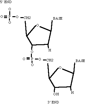
8) In the space below, draw the structure of one strand of a DNA dinucleotide.
You do not have to draw the bases, just indicate where they are located.
Do not use a circle and P for the phosphate(s). Label the 5' and 3' ends.

6 pts.
9) What was the karyotype of the Japanese woman that was infertile and we
read about? What mutation caused her to be female?
She had the normal karyotype of a man with one X and
one Y chromosome.
The mutation was a nonsense one where a stop codon appeared in the middle
of her SRY gene so that she could not produce the SRY protein and thus not
develop into a male. Therefore she was a female.
12 pts.
10) Explain the role of these components in the production of mRNA
a) the promoter
This is the DNA sequence just a few nucleotides upstream
of the start transcription site of a gene where transcription factors will
bind before the gene can be transcribed.
b) spliceosome
This is a mixture of RNAs and proteins that forms
a complex within the nucleus. Its function is to splice out the introns
of hnRNA and to splice together the remaining exons in the formation of
an mRNA.
c) introns and exons
Introns are parts of the gene that get transcribed
but will not be translated since they are spliced out of the hnRNA before
the mRNA is exported to the cytoplasm. Exons are the parts of a gene that
are retained in the mRNA and will be seen by the ribosome (most of the exons
will be translated).
8 pts.
11) How much energy is required to translate a protein that is 10 amino
acids long? Include in your analysis the amount of energy to add the amino
acids to the tRNAs, and all of the steps in translation. Show your
work for any partial credit.
10 ATP to couple amino acids to 10 tRNAs.
1 GTP to get the first amino acid (met) in the ribosome.
18 GTPs for the assembly of the next 9 amino acids (2 for each amino acid).
TOTALS: 10 ATP and 19 GTP.
4 pts.
12) What is a signal sequence and what is its function?
A signal sequence is a combination of amino acids
on the first (amino) end of a protein as it is being made. If the right
combination is there, the protein has a signal sequence and translation
of the protein will be completed on the rER. If the newly emerging protein
does not have a signal sequence, then the entire protein will be made on
a free floating ribosome and not the rER.
8 pts.
13) What is the specific genetic cause (at both the DNA and protein level)
for sickle cell? A complete answer will include not only a description of
what changes are present but also why this leads to the phenotype.
There is a single nucleotide change in a codon that
leads to a missense mutation. Specifically, in the beta chain of hemoglobin,
at amino acid number 6, there is a swap of a fully charged amino acid for
an hydrophobic amino acid. This causes the beta chain, and the entire tetramer,
to form a crystal structure along with all the other mutant proteins whenever
there is a low oxygen condition. These crystals lead to malformations of
the RBCs, clotting etc.
4 pts.
14) In a breakthrough experiment, Sato and Sato developed an assay to measure
what was wrong with CF cells. What was the conclusion of their research?
They concluded that CF cells were not able to secrete
Cl- in response to cAMP. This failure is not due to a lack of functional
cAMP-dependent protein kinase, but because this kinase could not phosphorylate
its substrate properly in mutant cells.
12 pts.
15) A family farm in New Zealand produced 450 sheep and 650 goats. The results
are shown below for the two herds. Remember that different species organize
their genes differently so don't compare apples and oranges (or in this
case kids and lambs).
a) Determine if the traits for each species are linked or not, and explain your answer.
b) If there is any linkage, determine the distance (in map units) between the loci.

a) For goats, there is no linkage since each of
the four possible genotypes is equally represented.
For sheep however, there is linkage since the four genotypes are not equally
represented.
b) For sheep, there are 32 map units between the two alleles. 144 / 450 = .32 or 32% recombination freq.