This web page was produced as an assignment for an undergraduate course at Davidson College.
Cellular Immune Response
The term cell-mediated immunity refers to the antigen-specific action of T cells. T cells, so named because they mature in the thymus, can be categorized as CD4 or CD8 cells, depending on which cell surface marker they express. All T cells express T cell receptors (TCRs), but different T cells recognize different antigens because of the high variability in antigen-binding region of TCRs, allowing the immune system to recognize and fight off many different pathogens. To become activated against a pathogen, a T cell needs two signals from an antigen-presenting cell (APC), such as a macrophage or a dendritic cell. The first signal is the interaction of the TCR and the peptide:MHC (major histocompatibility complex) complex found on the APC. The second signal is the interaction of costimulatory molecules B7, found on the APC, and CD28, found on the T cell, although CD8 cells can also receive a signal through a Fas:FasL interaction with a virally infected cell. The two types of T cells interact with different MHCs; CD4 T cells interact with MHC-II and CD8 T cells interact with MHC-I. MHC-II molecules present extracellular antigens that the cell has phagocytosed and processed, while MHC-I molecules present intracellular antigen, such as viral or intracellular bacterial particles.
Once a CD8 cell is activated, it becomes a cytotoxic T cell, or a CTL. CTLs kill infected cells by inducing apoptosis. CTLs form tight junctions, called super molecular adhesion complexes (SMACs) with infected cells, then release substances such as perforin, granzymes, and granulysin to induce apoptosis in the infected cell. And the SMAC prevents these substances from affecting neighboring cells. CTLs also secrete cytokines with various functions. For example, INF-γ prevents viruses from replicating, causes upregulation of MHC-I by other host cells, and enhances the antigen-presenting and effector functions of macrophages. Other cytokines cause a local inflammatory response, which includes increasing the permeability of blood vessels, allowing fluid to lead into the surrounding tissue.
Some CD4 cells are also involved in the cellular immune response. CD4 cells can be further divided into two groups: Th1 or Th2 cells. Th2 cells are mostly only involved in the humoral response, while Th1 cells are involved in both the cell-mediated and humoral immune response. Once a Th1 cell is activated, it participates in the cellular immune response primarily by activating macrophges. Th1 cells can produce both signals necessary for macrophage activation: IFN-γ and CD40 ligand, a cell-surface protein that binds to CD40 on the macrophage surface. Th1 are also involved in the direct killing of infected cells by the secretion of TNF-β and the upregulation of the surface protein Fas ligand. Th1 also recruit macrophages to the site of infection (Janeway et al. 2005). In a normal immune response, approximately 1/10,000 T cells are activated (Todar 2002).
While S. pyogenes is normally susceptible to cell-mediated immune defenses, in rare cases it causes a large immune response that is damaging or fatal to the infected individual. This occurs when secreted exotoxins act as superantigens (Todar 2002). Superantigens are the only protein antigens that can activate T cells without being presented on MHC-II (Janeway et al. 2005). Superantigens cause nonspecific TCR:MHC-II interactions, resulting in the stimulation of up to 20% of an individual's T cells. These stimulated T cells secrete a large amount of cytokines, causing all blood vessels to become more permeable. This results in decreased blood volume, which causes collapse of small vessels and clotting in others. Eventually, this failure of the circulatory system causes organ failure and death (Janeway et al. 2005). Out of the approximately 9,000 cases of invasive GAS infection that occurred in the U.S. in 2002, less than 6% resulted in streptococcal toxic shock syndrom (STSS), although 45% of those cases resulted in death (Center 2005).
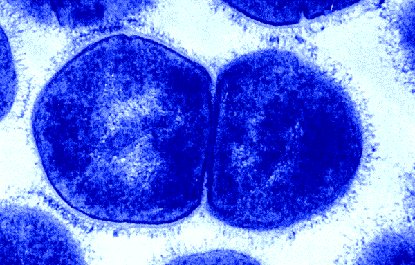
Figure 5. Two S. pyogenes bacteria. Electron micrograph by Maria Fazio and Vincent A. Fischetti, Ph.D. The Laboratory of Bacterial Pathogenesis and Immunology, Rockefeller University. Copyright 1995. Used with permission.
Return to S. pyogenes homepage
Davidson College Biology Home Page
Send any comments or concerns to beenglish@davidson.edu
© Copyright 2007 Department of Biology, Davidson College, Davidson, NC 28035