**This webpage was produced as an assignment for an undergraduate course at Davidson College**
Humoral Immune Response
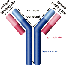
According to Whitley and Miller (2001), the adaptive immune system quickly responds to HSV infection with a humoral response, which involves neutralization, opsonization, and complement activation (Janeway 2005). B cells that differentiate into plasma-secreting cells produce antibodies that can bind HSV epitopes in the antigen binding site (see Figure 1). The predominant antibodies against HSV belong to the IgA isotype, and this type of antibody is secreted by plasma cells. IgA can be detected 3 days after infection, IgG1 and IgG3 are detected next, and finally IgM (Whitley and Miller 2001). Antibodies against gD and gB reduce the spread of HSV-1 through axonal transport, and this is one way that the immune system controls HSV infection (Mikloska et al. 1999). However, while antibodies produced by B cells can neutralize HSV, they cannot halt HSV replication or reactivation (Braun et al. 2006) . Studies have found high levels of antibody during viral reactivation (Rajcani and Durmanova 2006). This information about the inability of antibodies to stop HSV infection highlights the importance of the cell-mediated immune response and the challenges in developing a vaccine to prevent HSV infection.
The humoral response is dependent on the complement system as the complement system activates B lymphocytes and memory cell formation (Da Costa et al. 1999). When the complement receptors CD21/CD35 are blocked with antibody or soluble receptor, the humoral response is not fully functional, as CD35 binds to C4b and C3b. If the complement system is inhibited, CD35 is not activated by the complement components, and the humoral response is absent. Mice that had either C3, C4, or receptor CD21/CD35 deficiencies had a decreased antibody response the HSV (Da Costa et al. 1999).
One way that HSV evades the immune system is through the virion host shutoff (vhs) protein. vhs prevents MHC class II expression, which then inhibits T cell activation and subsequent T-cell dependent B cell activation (Smiley et al. 2004).

Figure 1. Antibody structure. Image from http://www.biology.arizona.edu/IMMUNOLOGY/tutorials/antibody/structure.html
Davidson College Biology Department
Contact jehodge@davidson.edu with questions or comments.