-
"This web page was produced as an assignment for an undergraduate course at Davidson College."
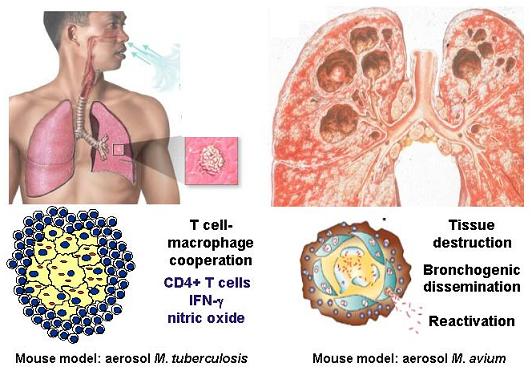
“Immunity to M. tuberculosis is regulated by T-cell and macrophage responses” (Barnes 1994).
A successful cellular immune response depends on Th1 activation.
Permission pending
Image provided with permission from: http://www.fz-borstel.de/en/research/ibm/mi/index.htm
Research Center Bostrel, Leibniz- Center for Medicine and Bioscience
αβ T-Cell Response
Both CD4+ and CD8 T+ cells play a significant role in the immune response against M. tuberculosis, as evidenced by knockout experiments of CD4+ and CD8+ T-cell which severely exacerbate the infection (Chan 1994). CD8+ cells are important because host cells other than macrophages which become infected such as parenchyma cells in the lung are not recognized by CD4+. Only CD8+ T-cells can effectively handle these other types of infected cells (Chan 1994).
Though both CD4+ and CD8+ T-cells play a role in immune response, CD4+ cells are more crucial to the success of the immune response. CD4+ cells are then further subdivided into Th1 and Th2, and the type of CD4+ response can dictate vastly different response to a tuberculosis infection (Chan 1994). Th1 cells produce INF-gamma, IL-2, and lymphotoxin. Th2 cells produce IL-4, IL-5, IL-6, and IL-10. Th1 cells increase antimicrobial activity of macrophages, where Th2 cells support B-cell proliferation and differentiation. Th1 cells help control the disease progression through INF-gamma, but Th2 cells hinders immune response through the production of IL-4 by inhibiting the Th1 growth (Barnes 1994). As a result, activation of a Th2 response generally leads to disease progression, whereas activation of a Th1 response leads to protection (WHO 2007)
M. tuberculosis replicates within vaculoes in macrophages, as a result MHC-II recognition of the pathogen via CD4+ cells is important. CD4+ Th1 cells release substantial amount of INF-gamma. INF-gamma plays a crucial role in mounting the immune response to M. tuberculosis, because induces antimycobacterial activity in macrophages, specifically inducing NOS2 expression (Chan 1994). CD4 + T-cells also have the capability to lyse phagocytes displaying mycobacteria on the MHC class II receptors. These cells lyse infected cells through perforin (bores a hole in the infected cells membrane) and granulysin (activates the capase cascade) (Flynn 2001). More effective phagocytes then hopefully take up exposed bacteria. The process of lysis and macrophage activation continues. However, target cell lysis causes host cell damage in the form of tissue and organ damage. Therefore, lysis of M. tuberculosis infected cells can be detrimental or beneficial depending on the circumstances (Chan 1994).
CD8+ cells have two effector functions in the immune response to tuberculosis: lysis of infected cells and production of cytokines, INF-gamma. CD8+ cells can lyse infected cells by perforin/granulysin or Fas/FasL pathway. Some M. tuberculosis antigens have been presented on MHC-I molecules, but the mechanism by which these antigens gain access to MHC-I is not understood (Flynn 2001).
gamma delta T-cell Response
In addition to αβ T-cells, gamma delta T-cells have also been shown to respond in the mycobacterium infections. In vitro, gamma delta T-cells are activated by mycobacteria and have been shown to acculmulate at the site of infection. gamma delta T cells have been reported to secrete INF-gamma and are considered part of the early immune response. Whereas CD4+ cells are vital for protective immunity and memory (Tsukaguchi 1995).
This site was created as partial fulfillment of requirments for Biology 307 at Davidson College in the Spring Semster of 2007.
Davidson Home Davidson Biology Home
Questions and comments should be directed to Dr.Sarafova or to the site creator Emily Rivard