Life Cylce
Baccillus anthracis is the bacteria that causes anthrax, an often fatal infection of the skin, respiratory system (inhalational anthrax), or gastrointestinal system (1). The disease affects almost any animal, but those most susceptible are large herbivores (1,2,3). Humans are affected through their interactions with these herbivores products derived from their corpses; inhalational anthrax is sometimes referred to as woolsorter’s disease due to the prevalence of the infection among those who handle wool (2). While theoretically possible, there have been no cases of human to human anthrax transmission in the literature (2).
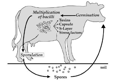
Basic anthrax lifecycle. See source 4
Cutaneous anthrax is characterized by painless, black-ended necroses at the site of infection, along with edema of the surrounding tissues. It is the most common form of anthrax infection, accounting for 95% of reported cases in the United States. It is generally not a life threatening condition (1).
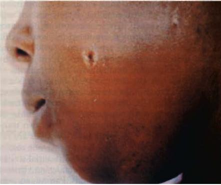
Cutaneous anthrax lesion (See source 1).
Gastrointestinal anthrax is more serious, although it has never been reported in the United States. The main symptoms involve bleeding into the stool and vomit, along with abdominal pain. Death can occur from perforation of the intestines or accumulation of anthrax toxins. However, if a patient survives, symptoms subside within two weeks (1).
Inhalational anthrax is rare, but extremely dangerous. In a 1979 outbreak in Sverdlovsk, USSR, only 20% of those suffering from inhalational anthrax survived. From the onset of initial symptoms, which resemble an upper respiratory tract infection, the mean time of death is only three days (1).
Morphology
B. anthracis is a rod-shaped, gram-positive bacteria. It is square-ended and non-motile, with the cells often occurring in vivo in long chains that resemble bamboo (2). The first part of the bacteria to interact with the host, when it is in its spore form, is the exosporium. It is made mostly of protein, with lipid and carbohydrate components (4). While the function of the exosporium is unknown, it appears to have pili that seem to enhance spore attachment to surfaces or ligands.
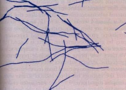
Photomicrograph of B. anthracis (see source 1).
B. anthracis also has a unique capsule which is considered to be a major contributor to its virulence. The capsule enhances the bacteria’s ability to evade host defenses, as well as inducing septicemia. It is constructed of a polymer of g-D-glutamate, synthesized by a enymatic complex that is part of the B. anthracis membrane. The enymatic complex is encoded on the pXO2 plasmid (4).
Finally, the S-layer is the final bacterial layer covering the peptidoglycan. The S-layer completely covers the cell surface. In B. anthracis, the S-layer is constructed of two similarly sized proteins. The two proteins, surface array protein and extractable antigen 1, self-assemble on the protein surface. The S-layer is thought to aid the capsule in evading host defenses, though its functions are not yet completely understood (4).
Initial Infection and Activation of Vegetative State
Anthrax exists in both a vegetative and spore state. The spores are extremely resistant, and have been a popular subject of discussion in both the scientific literature and the mainstream media. These spores make a good starting point for a discussion of the pathogen’s lifecycle. Herbivores, especially those in daily contact with soil (3), become infected with the spores. The spores are elliptical in shape (4), and resistant to heat, drying, ultraviolet light, and gamma radiation (1). The spores do not divide, and have no measurable metabolism (1). Once ingested, spores get taken up by macrophages and carried to lymph nodes (4). The gerH locus, a tricistronic operon present in B. anthracis, helps initiate germination with help from cogerminants unwittingly provided by macrophages (7). The spores enter the vegetative state in the macrophages, lysing them, and eventually overwhelming the lymph node. The infection then enters the blood stream, where the infection can be as intense as 107-108 per milliliter of blood (1). Anthrax in these animals tends to be very severe, and those in the late stages of infection bleed through the nose, mouth, and bowel. The blood exposes the surrounding soil with vegetative bacteria, as does the corpse of the infected animal when it dies (2).
Sporulation
Anthrax spores form when vegetative bacteria is exposed to oxygen (2,5). Spore formation is an extremely taxing process, involving the expenditure of a large amount of energy as many gene that are silent in the vegetative state are activated. To further complicate matters, sporulation is usually initiated at a time in which nutrients are scarce (6). For this reason, sporulation is a tightly and elaborately controlled process. In all Bacilli, the process involves a phosphorelay with a two component signal transduction system. Sensor kinases are activated by environmental or metabolic stimuli autophosphorylate on a histidine residue. The phosphoryl group is transferred on an aspartate on Spo0F and a histidine on Spo0B to an aspartate on Spo0A, which is the master control element in the initiation of sporulation in anthracis bacillus (6).
Persistence in the Environment
Once sporulation occurs, the spores can lay dormant in the soil for several years, although evidence suggests that spores vegetate in soil under favorable conditions (3). The soil B. anthracis is most commonly found in is that in which water has stood long enough to kill grass. This soil has a high content of organic material. Evidence also suggests that B. anthracis is most stable in soil with a pH above 6.0 and an ambient temperature above 15.5o C (3). These spores either die, or are ingested by another host at which point the cycle repeats itself.