This site was created as an undergraduate course requirement at Davidson College
Trichophyton
Evasion of Immune Response Detection
It is believed that Trichophyton invades the upper epidermal layers by releasing serine proteases that aid in the penetration through the skin (Kaufman et al 2007). These proteases also hydrolyze the keratinocytes providing the necessary nutrients for the pathogen. Trichophyton uses long and short fibrils to anchor its arthroconidia to the upper stratum corneum, while using only shirt and thin fibrils to penetrate into the more densely packed inner layers (Kaufman et al and Wang et al 2006). Trichophyton is very unique in the manner that its cell wall component mannan is believed to have a dual function in that it acts as both an antigen and an inhibitor of the host immune system. It is unknown whether it is the whole mannan molecule that is necessary for both functions or just pieces are necessary (Dahl 1994). It is believed that different portions of the mannan molecule function differently, such as possibly the gylcoproteins aid in inhibition of the host immune system, while the peptide portion is recognized as the antigen (Dahl and Grando).
Phagocytosis was inhibited by the Trichophyton exoantigen mannan, which binds to either the mannose receptor or the beta-glucan receptor (Campos et al.). The mannan binds to the cell surface receptors of only the monocytes and in studies, after 4-6 hours of infection granules are present with the nuclei and cytoplasm of infected cells (Dahl and Grando; Dahl). The internalized mannan molecules travel and accumulate in the nucleus of the cell. It is believed that here is were they alter the gene transcription or potential translation of proteins in the cell (Grando et al. 1992). As stated earlier, 1/3 of cases are asymptomatic and in vitro, it takes 48 hours for infection of Trichophyton to be detected (DoctorFungus 2007). Since mannan interferes with protein production, then there will most likely be altered proteins on the cell surface for the cell a macrophage to detect. It is important to state that the mannan ingestion does not kill the cell, but alters its function. It is important to note that it is only predicted and not yet proven that RNA synthesis is interfered by infected keratinocytes (Dahl and Grando).
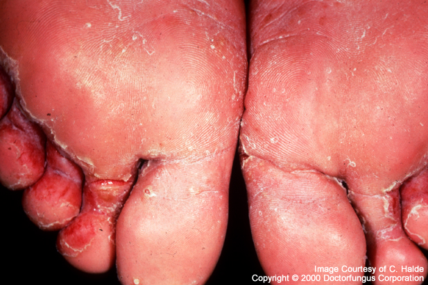
Figure 5. Foot sores from athlete's foot. Since the body is unable to eradicate the pathogen from the body completely sores like these often occur in the symptomatic patients. Even though the body uses natural defenses such as epidermal turnover and phagocytosis, one cannot rid the pathogen from the body (Permission pending http://www.doctorfungus.org/thefungi/trichophyton.htm).
Monocytes, not lymphocytes, can also uptake the conidia of Trichophyton and will reduce macrophage function (Grando et al.; Dahl and Grando). Ingestion of conidia caused necrosis of the macrophage within 8 hours of ingestion by growth and differentiation into more hyphae (Campos et al. 2006). Campos and his colleagues found that the infected macrophage was able to produce and secrete high levels of the pro-inflammatory cytokine TNF-alpha. Their tests also showed that macrophages infected with Trichophyton altered MHC II function. There was also noticeable down regulation of expression of the co-stimulatory molecules CD54 and CD80. In an attempt to try and combat the infected macrophage’s inflammatory response, Trichophyton induces production and release of the anti-inflammatory cytokine IL-10, which inhibits the Th1 response (Campos et al.). Thus the pathogen is mimicking the host’s Th2 response and not allowing it to attract more macrophages and other inflammatory cells (Janeway).
Another hypothesis to why the body cannot sustain a DTH response and a chronic infection persists, which has to do with the desensitizing the immune system, which Dahl and Grando called selective anergy. Because the infection usually occurs in conditions where the individual is continuously exposed to the pathogen due to dampness and mal hygiene, the body might stop producing a DTH response. It is believed that the mannan aids destruction of a DTH response by impeding T cell activation pathway by altering how the antigen, itself, is presented (Dahl and Grando).
Prevention & Treatment and Future Research
For more information, please refer to the
Davidson College Biology Home Page
E-mail me any comments or concerns
© Copyright 2007 Department of Biology, Davidson College, Davidson, NC 28035