This web page was created for an undergraduate molecular biology course at Davidson college.
Check out the pQE-30 UA Vector Map
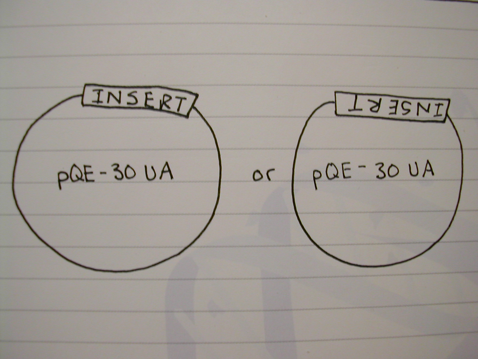
This simple diagram shows the problem of insert orientation. Due to the nature of sticky ends, our PCR insert could insert into the Plasmid in either of two orientations. One would put our IDH gene in the correct orientation, and this would lead to the desired fusion protein. However, the other orientation would lead to a non-functional protein. We must therefore screen the plasmid DNA of our cells to determine which insert orientation is present in their plasmids.
Diagram 1
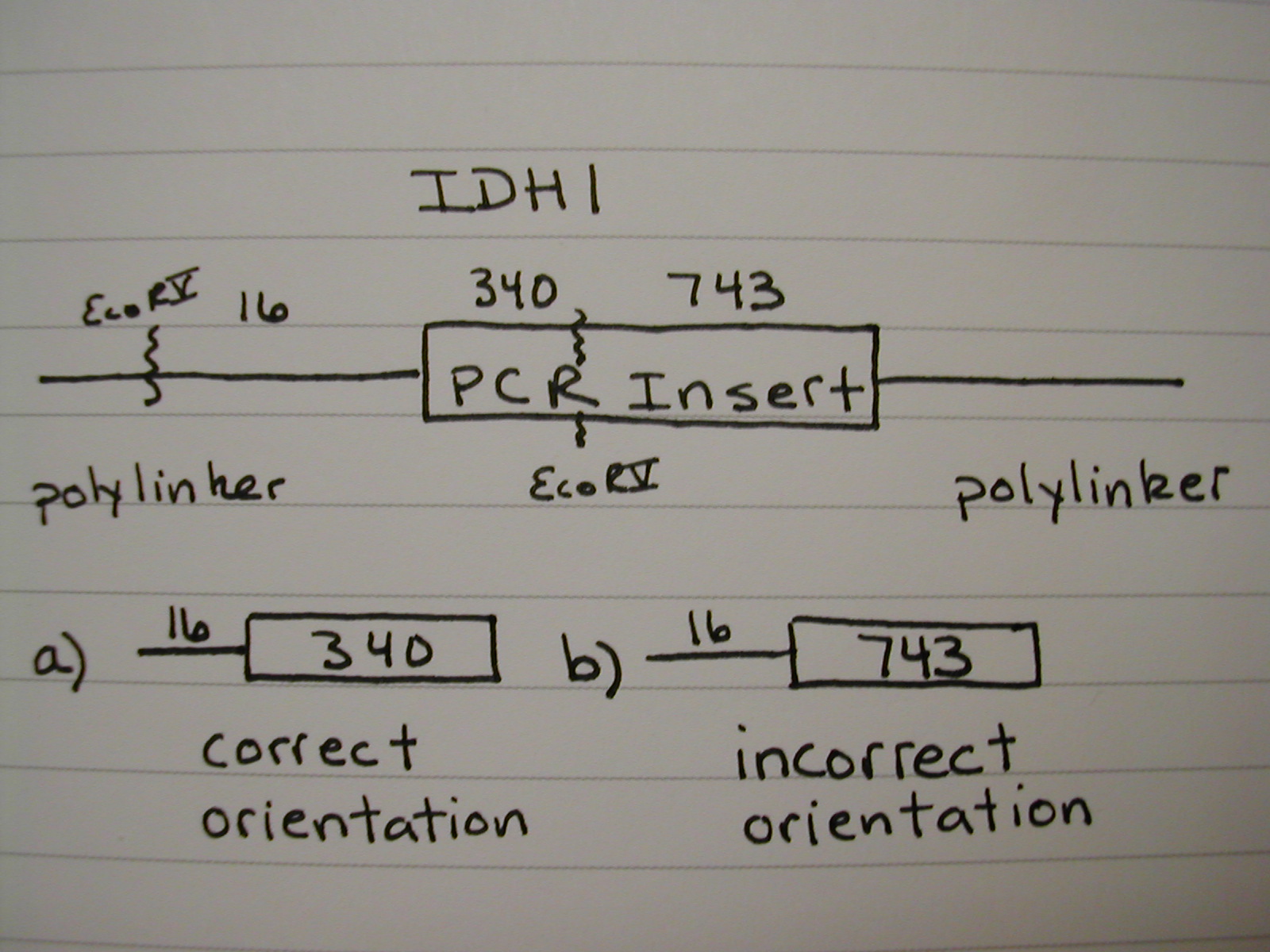
To test orientation we will use Restriction Enzymes and our knowledge of the IDH gene sequences. We can predict the length of the restriction fragments in both the correct and incorrect orientations. Diagram 2 shows a schematic representation of the restriction cuts and resulting fragments for IDH1 after being digested with Eco- RV. The insert is shown in the correct orientation. Note b) which shows the restriction fragment that would result had the PCR insert been inserted backwards. It is important to pick a restirction enzyme that cuts only once in the PCR insert in order to test the orientation of the insert. It should also be noted that this experiment is done one colony at a time. Within a colony all of the plasmids will be the same because E. coli are clonal.
Diagram 2
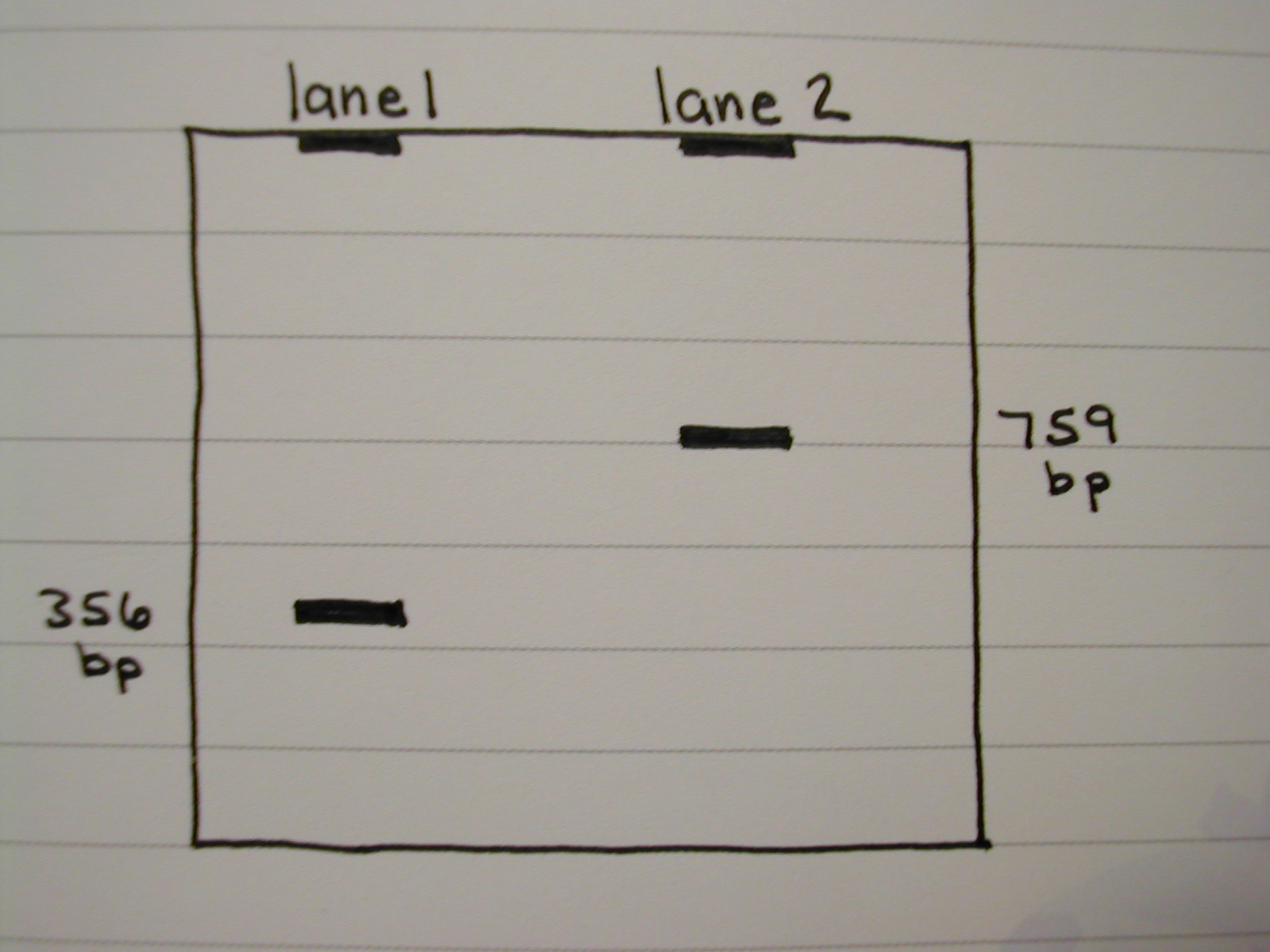
Once you predict your correct and incorrect band lengths, you digest the plasmid DNA from one colony with the selected restrictin enzyme and run the fragments on a gel. You then view the gel to be sure that there is a band at the predicted correct length and not a band at the predicted incorrect length. Diagram 3 shows only the restriction fragments (and not the large piece of plasmid DNA that would also be on the gel) we desired. Lane 1 is from a cell that took up a plasmid with correct orientation of IDH1, and lane 2 from a cell that took up a plasmid with incorrect orientation.
Diagram 3
Contact Kevin James with comments or questions.