All fluorescent dyes emit light of one wave length (e.g. green) after they have absorbed light of another wave length (e.g. blue). However, if a very high intensity blue light is delivered to the dye, the dye will "photobleach" meaning that the high intensity light has rendered the dye unable to fluoresce.
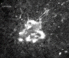
This phenomenon has lead to an interesting method called Fluorescence Recovery After Photobleaching (FRAP). The idea behind this method is to use FRAP to measure the ability of a molecule to move around over time. To do this, a fluorophore must be covalently attached to your favorite molecule (i.e. protein, lipid, carbohydrate). Using a epifluorescence microscope, visualize the fluorescently tagged molecule using a low light intensity (figure 1).
Figure 1. Monitoring the fluorescence before photobleaching.
Using a microscope allows the researcher to focus the light onto a small subset of the fluorescent molecules. Once the starting level of fluorescence has been determined for this small subset of molecules, flash a very intense light onto these same molecules. If all goes well, close to 100% of the fluorescent molecules will be photobleached and when the molecules are monitored again, the area will appear black due to the loss of fluorescence (figure 2).
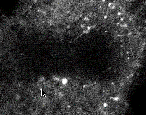
Figure 2. Monitoring the fluorescence after photobleaching.
Now there should be a black area filled with photobleached molecules surrounded by fluorescently tagged molecules that have not been photobleached. If these molecules are able to diffuse, they will. As they diffuse, the photobleached molecules and the fluorescent molecules will begin to mix. This will cause the fluorescent area to become a little less bright, but that is difficult to measure. However, the blackened area will gradually increase in brightness as fluorescent molecules migrate into this area (figure 3).
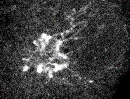
Figure 3. Monitoring the recovery of fluorescence after photobleaching.
Two parameters can be determined as the fluorescence returns to this photobleached area:
The raw data are collected by a light sensor which measures the light through the microscope. These data are displayed on a computer screen and by the end of the experiment, a graph will be produced that looks similar to figure 4.
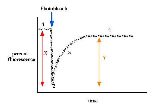
Figure 4. Graphical presentation of data collected during a FRAP experiment. A baseline of fluorescence is collected (1) before the photobleaching occurs (arrow) so that the amount of fluorescence is reduced significantly (2). Over time, the amount of fluorescence in the photobleached area increases as unbleached molecules diffuse into this area (3). Later, there is a stabilization of the amount of fluorescence recovery (4) and a flat line is obtained. The percent recovery uses the formula: (Y/ X) x 100 = % recovery. In the diagram, the percentage of fluorescence lost due to photobleaching is X and the amount of fluorescence that returned to the bleached area is Y. In practice, the percent recovery almost never reaches 100%. The lateral mobility is determined by the slope of the curve (3). The steeper the curve, the faster the recovery and therefore, the more mobile the molecules.
FRAP was used to help define the fluid mosaic model of cell membranes. It can also be used to determine if a protein is able to move within a membrane (high percent recovery with a fast mobility), or whether it is tethered other structural components of the cell (low percent recovery with a slow mobility). What would you predict for the percent recovery and lateral mobility for large multi-subunit integral membrane proteins that are not anchored to the cytoskeleton?