Results
from MacDNAsis
Open
Reading Frame and Molecular Weight
Hydrophobicity
Antigenicity
Predicted
Secondary Structure
Multiple
Sequence Alignment
Phylogenetic
Tree
View
3D Structure
This web page was produced as an assignment for an undergraduate
course at Davidson College
Return
to Jessica's Home Page
Molecular Biology Home Page
jereynoldskenneally@davidson.edu
Largest
Open Reading Frame
cDNA: Mus
Musculus
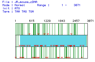
This figure represents the three possible reading frames for the Mus
Musculus cDNA
sequence. The largest open reading frame occurs at nucleotide
188 and ends at 3025.
View DNA sequences
from other species
Molecular Weight:
105569.01 Daltons
Predicted by MacDNAsis
Return to
top of the page
Hydrophobicity
Return to
top of the page
Kyte and Doolittle Plot
This protein
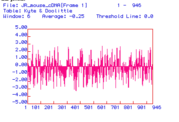
Any portion of this figure that reaches over 1.8 is a possible transmembrane
domain. There are many such domains for hexokinase. It
is therefore possible
that hexokinase spans a membrane. However I have not seen this
fact stated in
any literature I have reviewed on hexokinase. Another possibility
is that these
hydrophobic regions are necessary for the various interactions between
domains
of this molecular that make up it's two lobes.
View 3D structure
Antigenicity
Return to
top of the page
Hopp and Woods plot
would be a good portion
of the protein to use to generate a monoclonal antibody
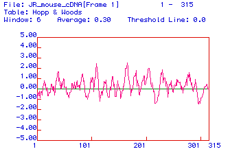
In this figure any portion of the graph which reaches 2 is a good candidate
for creating a monoclonal antibody to hexokinase. Therefore the
residues
near 110 and 180 have a sufficient amount of hydrophilicity to generate
a
good antibody.
Secondary
Structure
Return to
top of the page
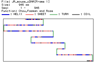
This figure shows the secondary structure of hexokinase. There
are many turns,
helicies, sheets and coils. This protein has a very complex secondary
structure.
While it is difficult to interpret exactly how this secondary structure
relates to the
3D image, it seems that this complex secondary structure could lead
to the globular
structure of hexokinase.
View
3D Structure
Multiple
Sequence Alignment
View DNA
sequences from other species
OR
Return to
top of the page
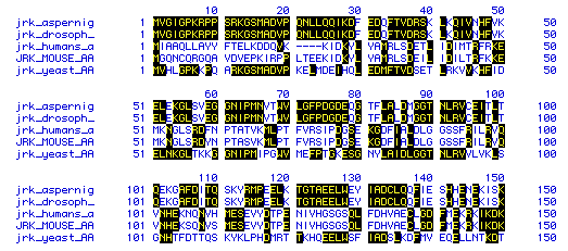
This figure shows a portion of the amino acid sequence from five species:
Mus
musculusHomo sapianS.cerevisiae
Drosophila
melanogasterAspergillus niger.
Five spaces had to be added
to humans to align that sequence with
the mouse sequence.
Phylogenetic
Tree
View DNA
sequences from other species
OR
Return to
top of the page
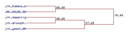
This figure shows the amount of conservation in the amino acid sequences
from the five species from above.
Drosophila
melanogasterand Aspergillus nigerare very closely related
as are the human and mouse
sequences. Yeast is closer
to Drosophila melanogasterand Aspergillus
niger than to the human and mouse.
Overall, hexokinase does not seem
very conserved.