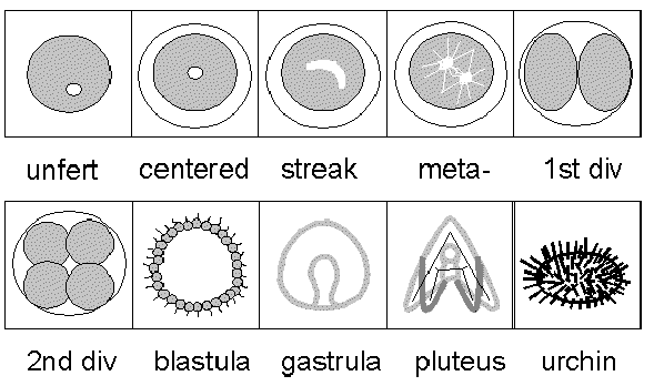
Cleavage Stage Embryos
Most introductory biology textbooks will cover aspects of sea urchin development and you might want to read that portion of your old textbook. The key topics used in Genomics, Proteomics & Bioinformatics are blastula and gastrula formation. Let's take a quick look so you will understand the timing and terminology under investigation in Eric Davidson's lab where he has uncovered the genomic circuitry of a single gene.
Cleavage Stage Embryos
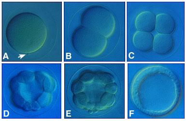
Figure 1. L. variegatus zygotes, viewed from the side. A. 1-cell zygote. The fertilization envelope is visible as a large "halo" around the embryo. The arrow points to the site of sperm penetration. B. 2-cell C. 8-cell D. 16-cell E. 32-cell F. Hatched blastula. (from worms.zoology.wisc.edu/urchins/SUcleavage_stages.html)
Click here to see Quicktime movie of cleavage.
Blastula Stage
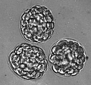
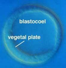
Figure 2. Left: Unhatched blastulae. Right: A L. variegatus hatched blastula, showing the thickened vegetal plate and obvious blastocoel. (from worms.zoology.wisc.edu/urchins/SUcleavage_blastula.html)
Click here to see animated gif
Click here to see a 3D animated gif
Gastrula Stage
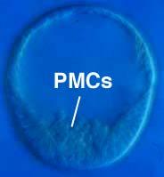
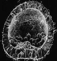
Figure 3. Left: L. variegatus mesenchyme blastula, viewed from the side, showing ingressing primary, or skeletogenic, mesenchyme cells (PMCs). Right: A L. variegatus mesenchyme blastula processed for scanning electron microscopy and fractured through the vegetal plate to show the process of ingression (from worms.zoology.wisc.edu/urchins/SUgast_ingression.html)
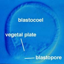
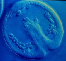
Figure 4. Left: L. variegatus early gastrula, viewed from the side. (from worms.zoology.wisc.edu/urchins/SUgast_primary.html) Right: L. variegatus late gastrula, viewed from the ventral (oral) side. (from worms.zoology.wisc.edu/urchins/SUgast_intro.html)
See a really good animated gif of gastrulation.
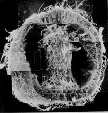
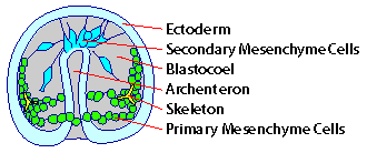
Figure 5. Left: L. variegatus late gastrula processed for scanning electron microscopy. Right: Cartoon of sea urchin gastrulat with some key parts labeled. (from www.luc.edu/depts/biology/dev/seaurgas.htm)
Overview of Development and Cell Fate Maps
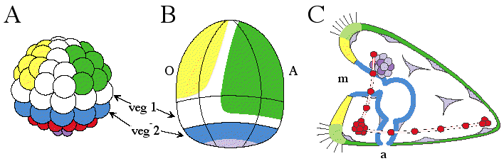
Figure 6. Developmental stages and fate maps of sea urchin S. purpuratus embryogenesis. (A) 60-cell stage embryo undergoing rapid cell division. Eight large micromeres (shown in red) will become mesenchymal cells destined to form the future skeleton. Beneath them are the four small micromeres (shown in purple). Though their progeny will contribute exclusively to the coelomic pouches, their actual state of specification at 6th cleavage is not known. The eight blastomeres of the veg2 ring are also specified and their progeny will express vegetal plate marker genes. The veg1 domain, here shown in white, is not yet specified, since later its progeny will be allocated into one of three different fates, nor are the lateral ectodermal domains on either side of the embryo (white arch of cells over the top) yet specified. Some progeny of these blastomeres will form the boundary regions between oral (yellow) and aboral (green) ectoderm. The polar clones of the aboral ectoderm territory are already specified, and these clones will soon begin to express aboral markers. The polar clones of the oral ectoderm territory may also be specified. (B) External lateral view of 24 hour old blastula composed of about 500 cells. Regions where specifications have still not yet occurred are shown in white. (C) Lateral view of final state of specification in early pluteus-stage larva, about 1500 cells (65 hours post-fertilization). (with permission from Eric Davidson; from Davidson et al. 1998. Development. Vol. 125: 3269 - 3290. Figure 2 on page 3271)
(with permission from SUE <http://www.stanford.edu/group/Urchin/dev.htm>)
2D Animated GIF of development to pluteus-stage larva.
Two 3D Animated GIFs of development to pluteus-stage larva.
Interactive Fate Map for Cells after Six Rounds of Division
Tutorial on Cleavage/Blastula Stage Sea Urchins
Tutorial on Gastrula Stage Sea Urchins
Images and Explanation of How Cells Invaginate During Gastrulation
Movie of Elongating Archenteron
Overview of Entire Sea Urchin Life History
Sea Urchin Development (SUE) comprehensive web site
![]()
![]()
© Copyright 2001 Department of Biology, Davidson College, Davidson, NC 28036
Send comments, questions, and suggestions to: macampbell@davidson.edu