
Figure 1. Structure of Cyclosporin A (Friedman 1991).
Immunology Protein Page
Nuclear Factor of Activated T Cells
Timothy S. Deeb
Immunology
In activated T cells, "signaling via T-cell receptor-associated tyrosine kinases" induces the synthesis of the "nuclear component of the nuclear factor of activated T cells (NFATn)" (Janeway and Travers 1994). In T cells, NFATc is an inactive, phosphorylated component of NFAT found in the cytosol which contains a nuclear localization signal. An increase in intracellular calcium ion concentration activates calcineurin, a "cytoplasmic-calcium-calmodulin-dependent serine/ threonine phosphatase" ( Abbas, Lichtman, and Pober 1997). Calcium activated calcineurin dephosphorylates NFATc and reveals a nuclear localization signal which allows NFATc to migrate to the nucleus. In the nucleus, NFATc binds to NFATn to form active NFAT. The activated transcription factor, NFAT, forms a complex with another transcription factor, AP-1 (a dimer composed of Fos and Jun proteins), and binds to the enhancer region of the cytokine gene. This NFATAP-1 complex and other transcription factors augment IL-2 gene transcription according to Abbas, Lichtman, and Pober. They suggest this type of NFATAP-1 cooperation "has been demonstrated in the transcriptional regulation of both the IL-2 and IL-4 genes" (Abbas, Lichtman, and Pober 1997).
"T cells express low levels of calcineurin, so they are more sensitive to inhibition of this pathway than are many other cell types," which results in increased selectivity (Janeway et al 1999). The important role of NFAT "in T-cell activation is illustrated by the effects of the selective inhibitors of NFAT called cyclosporin A and FK506 (tacrolimus)" (Janeway et al 1999). These immunosuppressive drugs inhibit allograft rejection in organ transplant patients. Cyclosporin A (see figure 1) enters the cytoplasm and binds to the intracellular immunophilin cyclophilin and then forms a complex with calcineurin, which prevents calcium from binding to and activating calcineurin.
Figure 1. Structure of Cyclosporin A (Friedman 1991).
FK506 (see figure 2) enters the cytoplasm and binds to the intracellular protein FK506 binding protein (FKBP) (Abbas, Lichtman, and Pober 1997). In T lymphocytes, FK506 binds to FKBP-12 and then forms a "complex of tacrolimus-FKBP-12, calcium, calmodulin and calcineurin," thus inhibiting the phosphatase activity of calcineurin (Burnham 2000).
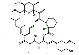
Figure 2. Structure of FK506 (Friedman 1991)
The cyclosporin A and tacrolimus complexes with calcineurin prevent
the calcium activation of calcineurin and in this way inhibit the activation
of NFATc by calcineurin. Because of the sensitivity of the T cells to the
selective inhibition of these drugs, allograft rejection is reduced.
Abbas, A.K., Lichtman, A.H., Pober, J.S. Cellular and molecular immunology. 3rd ed. Philadelphia: W.B. Saunders Co.: 1997 p 166-167.
Burnham, T.H, editor. Drug facts and comparisons. St. Louis: Facts and Comparisons: 2000 p. 1569.
Friedman, T. Molecular genetic medicine. Vol. I. SanDiego: Academic Press, Inc.: 1991 p. 148-149.
Janeway, C.A., Jr., Travers, p. Immuno biology. San Francisco: Current Biology Ltd.: 1994 p. 12:3-12:5.
Janeway, C.A., Jr., Travers, P., Walport, M., Capra, J.D. Immuno biology : the immune system in health and disease. New York: Elsevier Science Ltd./Garland Publishing: 1999 p.180.
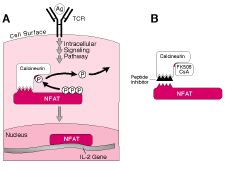
Figure 3. Illustration of a peptide that inhibits calcineurin from binding to NFAT. To learn about a new peptide that inhibits the signaling pathway of NFAT and that may be developed into a new immunosuppresssant, click the above image taken from the following publication: Peptide Points to Organ Preservation with Fewer Side Effects. (Permission to use this image has been granted by Robert Neal at HMS.)
![]()