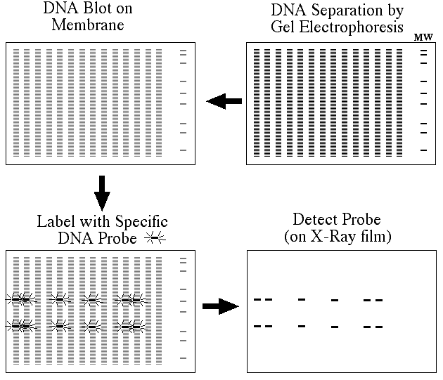
This is a brief overview of how a Southern blot (more formally called an DNA blot) is performed and what type of data you can obtain form one.

Figure 1. Southern blots allow investigators to determine the molecular weight of a restriction fragment and to measure relative amounts in different samples.
Procedure
1) DNA (genomic or other source) is digested with a restriction enzyme and separated by gel electrophoresis, usually an agarose gel. Because there are so many different restriction fragments on the gel, it usually appears as a smear rather than discrete bands. The DNA is denature into single strands by incubation with NaOH.
2) The DNA is transfered to a membrane which is a sheet of special blotting paper. The DNA fragements retain the same pattern of separation they had on the gel.
3) The blot is incubated with many copies of a probe which is single-stranded DNA. This probe will form base pairs with its complementary DNA sequence and bind to form a double-stranded DNA molecule. The probe cannot be seen but it is either radioactive or has an enzyme bound to it (e.g. alkaline phosphatase or horseradish peroxidase).
4) The location of the probe is revealed by incubating it with a colorless substrate that the attached enzyme converts to a colored product that can be seen or gives off light which will expose X-ray film. If the probe was labeled with radioactivity, it can expose X-ray film directly.
Below is an example of a real Southern blot used to detect the presence of a gene that was transformed into a mixed cell population. In this Southern blot, it is easy to determine which cells incorporated the gene and which ones did not.
Figure 2. The figure on the left shows a photograph of a 0.7% agarose gel that has 14 different samples loaded on it (plus molecular weight marker in the far right lane and a glowing ruler used for analysis of the results). Each sample of DNA has been digested with the same restriction enzyme (EcoRI). Notice that the DNA does not appear as a series of discrete bands but rather as a smear. The DNA was transferred to nitrocellulose and then probed with a radioactive fragment of DNA that was derived from the transformed gene. The figure on the right is a copy of the X-ray film and reveals which strains contain the target DNA and which ones do not.
© Copyright 2001 Department of Biology, Davidson College, Davidson, NC 28036
Send comments, questions, and suggestions to: macampbell@davidson.edu