This web page was produced as an assignment for an undergraduate course at Davidson College.
Humoral Immune Response
The term humoral immune response refers to immunological action of antigen-specific antibodies, which are secreted by activated B cells. Antibodies produce an immune response through three main mechanisms: neutralization, opsonization, and activation of the complement cascade. Neutralization simply refers to the process by which antibodies prevent virues and intracellular bacteria from entering host cells by binding to the proteins on the pathogen surface that are necessary for entry into host cells. Opsonization occurs when antibodies coat the surface of the pathogen, usually a bacteria, and enhance uptake of the pathogen by phagocytic cells. The complement cascade itself has two possible outcomes: opsonization by certain complement proteins, usually C3b, and direct killing of the pathogen by the formation of a pore in the plasma membrane by 10-16 molecules of the complement protein C9. There are five different isotypes of antibodies with different distributions and effector functions: IgM, IgD, IgG, IgA, and IgE. IgM is best at complement activation and transport across epithelium. It is also the first antibody produced when the host is infected with a pathogen for the first time. The function of IgD is not completely understood at this time, and it is never secreted. There are many different subtypes of IgG, and their effector functions vary to include, among others, opsonization and complement activation, and they often diffuse into extravascular sites. IgA is best at neutralization and is also is often transported across epithelium as a dimer. IgE is best at sensitization of mast cells and is often found in extravascular sites. (Janeway et al. 2005).
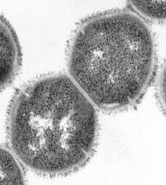
For most inidividuals, the humoral immune response to S. pyogenes is primarily based on IgG antibodies' binding to M protein on the bacterial surface. This binding causes opsonization, resulting in the phagocytosis and the eventual killing of the bacteria. Because the bacteria is often found on mucosal epithelial surfaces, S. pyogenes-specific IgA antibodies are also produced in the respiratory tract as protection against another streptococci infection. However, the protective properties of these antibodies are often negated by the extreme variability of the M protein (further discussed in the Evasion of the Immune System page).

Figure 4. Transmission electron micrograph of two S. pyogenes bacteria (20,000x). The outer filamentous material is mostly M proteins, and the clear region between the M proteins and the darker inside of the bacteria is the cell wall. . Electron micrograph by Maria Fazio and Vincent A. Fischetti, Ph.D. The Laboratory of Bacterial Pathogenesis and Immunology, Rockefeller University. Copyright 1995. Used with permission.
Return to S. pyogenes homepage
Davidson College Biology Home Page
Send any comments or concerns to beenglish@davidson.edu
© Copyright 2007 Department of Biology, Davidson College, Davidson, NC 28035