This web page was produced as an assignment for an undergraduate course at Davidson College.
Orthologs of Melanoma Inhibitory Activity (MIA)
This webpage identifies the orthologs of Melanoma Inhibitory Activity (MIA). Orthologs are genes evolved from a common ancestral gene. Through searches of the databases, Mouse Genome Informatics, BLAST, OMIM, Entrez, Swissprot, and PDB, the orthologs of MIA may be obtained. From analysis of the orthologs, information about the protein's possible phenotypes, viral and drug binding sites, similar proteins, and mutations can be obtained. Sequences of the protein that are maintained across species may be critical to the function of the protein, which explains why the sequence is evolutionarily stable. This webpage identifies some of these sequences.
The structure and sequence of MIA was originally found from human cells. The human MIA protein is 131 bp long and the amino acid sequence is MARSLVCLGV IILLSAFSGP GVRGGPMPKL ADRKLCADQE CSHPISMAVA LQDYMAPDCR FLTIHRGQVV YVFSKLKGRG RLFWGGSVQG DYYGDLAARL GYFPSSIVRE DQTLKPGKVD VKTDKWDFYC Q.
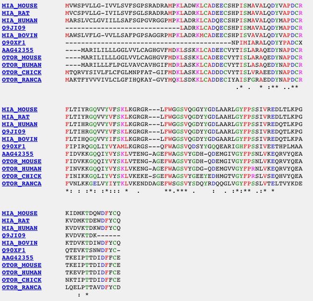
The MIA protein belongs in the family of proteins called MIA/OTOR. Species that contain the MIA or OTOR protein include mouse, chicken, bull frog, human, cow and rat. The proteins translated by the genes in this protein family share several amino acids:
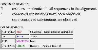
Fig. 1- Comparison of the amino acid sequences of the proteins in the family MIA/OTOR. Image courtesy http://pbil.univ-lyon1.fr/cgi-bin/acnuc-link-ac2aln?db=Hoverprot&query=Q61865
The Orthologs (by species):
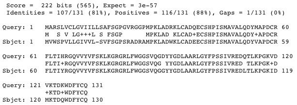
Mouse: From a BLASTp search:
Fig. 2- Comparison of the amino acid sequences of the human MIA protein and the mouse MIA protein. Image courtesy http://www.ncbi.nlm.nih.gov/BLAST/Blast.cgi#1816627
The mouse MIA protein is found in cartilage cells and has a phenotype identical to the phenotype of human MIA protein. Mice engineered with a mutation in the protein are still viable, but have some alteration in the structure of their cartilage.
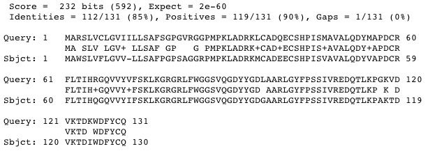
Rat: BLASTp search
Fig. 3- Comparison of amino acids in human MIA protein and rat MIA protein. Image courtesy http://www.ncbi.nlm.nih.gov/BLAST/Blast.cgi#13540667
The MIA protein in the rat functions in the development and maintenance of cartilage cells.
Cow: BLASTp search
Fig. 4- Comparison of amino acids in cow MIA protein and human MIA protein. Image courtesy http://www.ncbi.nlm.nih.gov/BLAST/Blast.cgi#27806849
The cow MIA protein functions in cartilage development and maintenance.
Conserved domains in other proteins:
An 18 residue loop found in the tertiary structure of MIA is homologous to the Src homology 3 (SH3) domains found in other proteins (from my favorite protein page):
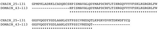
Fig. 5- The amino acids of the SH3 domain are lined up with the amino acids of MIA. Image courtesy http://us.expasy.org/cgi-bin/aligner
This domain was first discovered in several cytoplasmic tyrosine kinases. The domain is characterized by 5 or 6 tightly packed beta strands that act as two anti-paralell beta sheets. The function of this domain is to mediate the assembly of proteins by binding to polyproline helices (see my favorite protein page). Generally, the domain is only found once in a protein, but in some proteins the domain is found 3-4 times.
References:
SwissProt. 2005. Swiss-prot protein knowledgebase. <http://us.expasy.org/sprot/> Accessed 2005 8 Mar.
NCBI. 2005. National Center for Biotechnology Information. <http://www.ncbi.nih.gov/> Accessed 2005 8 Mar.
John Bunton's Molecular Biology Home Page
Questions or Comments: email me at jobunton@davidson.edu