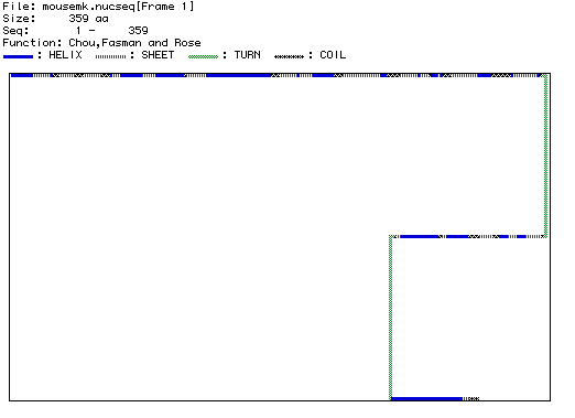
Predicted Secondary Structure of MAP kinase
This figure represents the secondary structure of MAP kinase. Blue segments represent alpha helices, while red-striped portions represent beta-pleated sheets. Green segments indicate turns and black segments with ** indicate coils. Overall the predicted secondary structure appears to be in agreement with the complex 3-D structure of MAP kinase. The predicted patterns of alternating alpha helices and beta-pleated sheets are supported by the more complex 3-D representation. In addition, the secondary structure correctly predicts a small number of widely spaced turns. Click here to compare the predicted secondary structure with a complex 3-D image of the structure of MAP kinase from Rattus norvegicus.
Back
to MacDNAsis Analysis of MAP kinase
Molecular Biology Home Page