DisplayPROFILETM-Restriction Fragment Differential Display PCR (RFDD-PCR)
DisplayPROFILETM RFDD is a method which uses the polymerase chain reaction (PCR) to make consistent and rapid gene expression profiles and to identify differentially expressed genes in various tissues and cells. To produce molecular snapshots of expressed genes, the displayPROFILETM technique involves digesting double-stranded cDNA with a restriction enzyme, ligating specific DNA adaptors to the cDNA fragments, and annealating specific primers onto the adaptors (Display Sytems Biotech). The cDNA fragments are then divided into 64 different PCR reactions. Each PCR reaction amplifies a different expression window, or a set of 400 or more cDNA fragments. Unlike standard differential display systems that use poly-A primed PCR amplification, RFDD-PCR uses the TaqI restriction enzyme and specific PCR primers for amplification (Display Systems Biotech). As a result of not relying on poly-A-primed PCR, this technique can be used for both eukaryotic and prokaryotic systems. The displayPROFILETM technique is important in the biotechnology world, for PCR methods can be used in the diagnosis of infectious diseases. In particular, this technique could possibly be used for cancer research since specific genes that are expressed or repressed in diseased versus healthy tissues can be easily identified and compared. Using RFDD-PCR, the genetic basis of specific cancers and new cancer therapies could be discovered.
The displayPROFILETM RFDD technique involves five major steps:
1. Double -stranded cDNA is synthesized from specific mRNA sequences
2. Double-stranded cDNA is digested with TaqI
restriction enzyme. Digestion with TaqI produces
sticky ends on the cDNA fragments.
3. Two specific DNA adaptors are ligated onto the sticky ends
of the cDNA fragments.
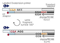
One of the adaptors is called the EP-adaptor, which consists of a 5'-overhang
and an
"extension-protection group" on its 3'-end. The purpose of the EP group
is to ensure that the
5'-overhang will not be filled in by 3' to 5' synthesis and that
cDNA fragments with EP adaptors at both
of its sticky ends will not be amplified during the polymerase chain reaction.
The second adaptor used
is called the standard adaptor. Only those fragments that have the EP adaptor
ligated onto one
of its ends and the standard adaptor ligated onto its other end are amplified
using PCR (Display
Systems Biotech).
4. Sixty-four high-strigency PCR
reactions are carried out with 0-extension 5'- primers and 64
different
displayPROBE 3'-primers. A 0-extension 5'-primer is annealed to the
EP adaptor on the
cDNA fragment, while one of the displayPROBE primers is annealed to the
junction between the cDNA
insert and the standard adaptor. Because a displayPROBE
primer consists of three bases
that extend into the insert cDNA, amplification of cDNA fragments with
a standard adaptor
ligated onto both ends is prevented (both ends would need the same three
bases to be amplified), and
each displayPROBE is specific for a certain cDNA sequence. Thus, in order
to amplify all fragment
variations, sixty-four displayPROBE primers are needed for each PCR reaction
(4^3=64) (Display
Systems Biotech).
When PCR is run, each of the 64 reactions amplifies an "expression window,"
which
consists of 400 or more fragments. Because each cDNA fragment can be oriented
in two different
orientations relative to the two adaptors, each amplified cDNA fragment
should be represented twice-
once in two different expression windows. See figure below:
5. The amplified gene fragments are separated on a gel
(Display Systems Biotech).
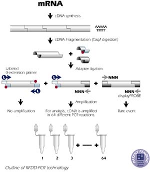
Figure 2. Illustration of how displayPROFILETM is
performed.
This image is courtesy of:<www.displaysystems.com>
Why is displayPROFILETMsuch
a great technique?
References:
Brown C. 1995. Homepage. <http://falcon.cc.ukans.edu/~jbrown/pcr.html>. Accessed 2000 Feb 20.
Display Systems Biotech. Homepage. <http://www.displaysystems.com>. Accessed 2000 Feb 20.
Display Systems Biotech. DisplayPROFILETM-Expression profiling using Restriction Fragment Differential Display (Brochure).
Macelis D, Roberts RJ. 2000 Feb 17. TaqI TypeII restriction enzyme.
<http://rebase.neb.com/rebase/enz/TaqI.html>.
Accessed 2000 Feb 20.
Please send comments to: casmith@davidson.edu