MacDNAsis Page
This page displays results from a MacDNAsis analysis of the human phospholipase C cDNA and amino acid sequence located on my genbank page. Also here is the MacDNAsis analysis of the homology between the respective amino acid sequences of the four other organisms listed on my genbank page and the amino acid sequence generated from largest open reading frams (ORF) in the human phospholipase C cDNA.
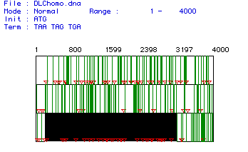
Phospholipase C cDNA Open Reading Frame.
Figure 1. Largest open reading frame of human phospholipase C cDNA.
MacDNAsis analysis of the human phospholipase C cDNA generated three open reading frames searches stacked here one on top of the other. The red triangles specify start codons, the green lines stop codons, and the black box indicates the largest open reading frame (ORF). The largest open reading frame, found in the third ORF search, lies between the 252nd and 3197th nucleotides. It produces a protein of 2945 bp. and a predicted molecular weight of 111.21073 kda. To view the original cDNA sequence from which the ORF was determined click here.
Hydropathy Plot of Phospholipase C.
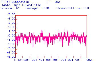
Figure 2. Hydropathy plot of the human phospholipase C amino acid sequence.
MacDNAsis analysis produced a hydropathy plot of the human phospholipase C protein. A hydropathy plot measures the hydrophobicity of the amino acids of a protein sequence. Peaks on the plot scaling to or beyond 2.00 suggest that those regions reside in a membrane. The plot generated shows some areas of hydrophobicity at and around amino acids 380 and 700. These peaks, however, are not high enough to be considered transmembrane regions. The lack of any transmembrane regions suggests that phospholipase C is not an integral protein.
Antigenicity plot of Phospholipase C.
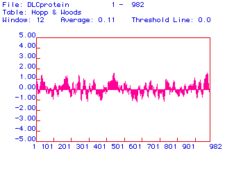
Figure 3. Antigenicity plot of the human phospholipasen C amino acid sequence.
MacDNAsis analysis produced an antigenicity plot of human phospholipase C. An antigenicity plot measures the hydrophilicity of the amino acids of a protein sequence. The greater the hydrophilicity the greater the cytoplasmic or extracellular exposure that particular portion of the protein experiences. A highly exposed epitope facilitates greater binding potential between itself and its corresponding antibody. Amino acid regions around 450 and 970 of the phospholipase C sequence show a high degree hydrophilicity. Portions of the protein such as these would be ideal epitope regions.
Predicted Secondary Structure of Phospholipase C.
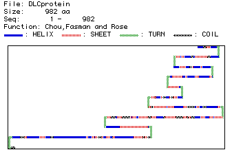
Firgure 4. Computer prediction of the secondary structure of human phospholipase C.
MacDNAsis analysis produced a prediction of the secondary structure of phospholipase C from the primary amino acid sequence. The blue portions indicate alpha helical regions, red portions indicate beta sheet regions, green portions indicate turns in the protein, and the gray portions indicate coiled regions. The predictated structure shows a great deal of turning throughout the protein and an extend alpha helical region toward the end of the protein. To view the three dimensional structure of phospholipase C click here.
Multiple Sequence Alignment of the Amino Acid Sequences of the Five Genbank Organisms.
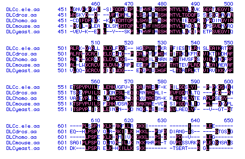
Figure 5. Multiple sequence alignment of the amino acid sequneces of the five genbank organisms.
MacDNAsis analysis produced a multiple sequence alignment of the amino acid sequences of the five genbank organisms. The black regions of the alignment indicate homology between organisms. There are certain areas within the alignment that show a high degree of homology. The portion seen above, however, details amino acid conservation of only 150 of the 982 total amino acids. The majority of the human phospholipase C protein shows a low degree of homology with the four other organisms. To view the orginal protein sequences of the five studied organisms click on any of the following: human; mouse; c.elegan; drosophila; or yeast.
Phylogenic Tree of the Amino Acid Sequences of the Five Genbank Organisms.
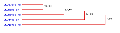
Figure 6. Phylogenic tree depicting the homology between the amino acid sequences of the five respective organisms.
MacDNAsis analysis produced a phylogenic tree detailing conservation between the amino acid sequences of the five genbank organisms. The tree shows little homology between the human phospholipase C protein and those of the four other organisms. The amino acid sequence from c. elegans had the highest degree of similarity at 15.5 per cent and yeast the lowest at 7.5 per cent. To view the original protein sequences of the five studied organisms click on any of the following: human; mouse; c.elegan; drosophila; or yeast.
To get to the genbank page click here.
To return to the DLC hompage click here.